Steidl Eike, Pilatus Ulrich, Hattingen Elke, Steinbach Joachim P, Zanella Friedhelm, Ronellenfitsch Michael W, Bähr Oliver
Dr. Senckenberg Institute of Neurooncology, Center for Neurology and Neurosurgery, Johann Wolfgang Goethe-University, Frankfurt/Main, Germany.
Institute of Neuroradiology, Center for Neurology and Neurosurgery, Johann Wolfgang Goethe-University, Frankfurt/Main, Germany.
PLoS One. 2016 Dec 29;11(12):e0168113. doi: 10.1371/journal.pone.0168113. eCollection 2016.
Antiangiogenic treatment of glioblastomas with Bevacizumab lacks predictive markers. Myoinositol (MI) is an organic osmolyte, with intracellular concentration changes depending on the extracellular osmolality. Since Bevacizumab markedly reduces tumor edema and influences the tumor microenvironment, we investigated whether the MI concentration in the tumor changes during therapy.
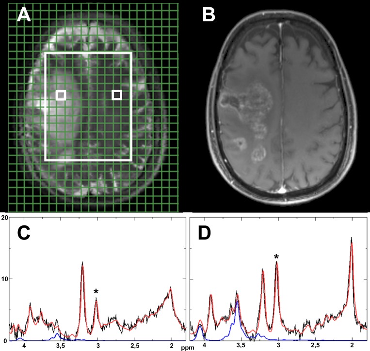
We used 1H-magnetic resonance spectroscopy to measure the MI concentrations in the tumor and contralateral control tissue of 39 prospectively recruited patients with recurrent glioblastomas before and 8-12 weeks after starting therapy. 30 patients received Bevacizumab and 9 patients were treated with CCNU/VM26 as control. We performed a survival analysis to evaluate MI as a predictive biomarker for Bevacizumab therapy.
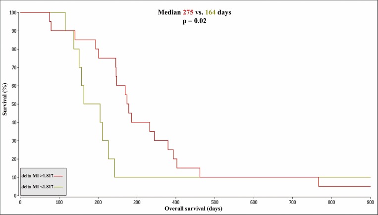
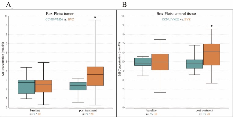
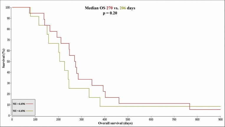
MI concentrations increased significantly during Bevacizumab therapy in tumor (p < .001) and control tissue (p = .001), but not during CCNU/VM26 treatment. For the Bevacizumab cohort, higher MI concentrations in the control tissue at baseline (p = .021) and higher differences between control and tumor tissue (delta MI, p = .011) were associated with longer survival. A Kaplan-Meier analysis showed a median OS of 164 days for patients with a deltaMI < 1,817 mmol/l and 275 days for patients with a deltaMI > 1,817 mmol/l. No differences were observed for the relative changes or the post treatment concentrations. Additionally calculated creatine concentrations showed no differences in between subgroups or between pre and post treatment measurements.
Our data suggest that recurrent glioblastoma shows a strong metabolic reaction to Bevacizumab. Further, our results support the hypothesis that MI might be a marker for early tumor cell invasion. Pre-therapeutic MI concentrations are predictive of overall survival in patients with recurrent glioblastoma treated with Bevacizumab.
使用贝伐单抗对胶质母细胞瘤进行抗血管生成治疗缺乏预测标志物。肌醇(MI)是一种有机渗透质,其细胞内浓度变化取决于细胞外渗透压。由于贝伐单抗可显著减轻肿瘤水肿并影响肿瘤微环境,我们研究了治疗期间肿瘤中的肌醇浓度是否发生变化。
我们使用氢磁共振波谱法测量了39例前瞻性招募的复发性胶质母细胞瘤患者在开始治疗前以及治疗8 - 12周后的肿瘤和对侧对照组织中的肌醇浓度。30例患者接受贝伐单抗治疗,9例患者接受洛莫司汀/替尼泊苷作为对照治疗。我们进行了生存分析,以评估肌醇作为贝伐单抗治疗的预测生物标志物。
在贝伐单抗治疗期间,肿瘤(p <.001)和对照组织(p =.001)中的肌醇浓度显著增加,但在洛莫司汀/替尼泊苷治疗期间未增加。对于贝伐单抗队列,基线时对照组织中较高的肌醇浓度(p =.021)以及对照组织与肿瘤组织之间较高的差异(肌醇差值,p =.011)与更长的生存期相关。Kaplan - Meier分析显示,肌醇差值<1,817 mmol/l的患者中位总生存期为164天,肌醇差值>1,817 mmol/l的患者为275天。相对变化或治疗后浓度未观察到差异。另外计算的肌酸浓度在亚组之间或治疗前和治疗后测量之间没有差异。
我们的数据表明,复发性胶质母细胞瘤对贝伐单抗表现出强烈的代谢反应。此外,我们的结果支持肌醇可能是早期肿瘤细胞侵袭标志物的假设。治疗前的肌醇浓度可预测接受贝伐单抗治疗的复发性胶质母细胞瘤患者的总生存期。