Torok Szilvia, Rezeli Melinda, Kelemen Olga, Vegvari Akos, Watanabe Kenichi, Sugihara Yutaka, Tisza Anna, Marton Timea, Kovacs Ildiko, Tovari Jozsef, Laszlo Viktoria, Helbich Thomas H, Hegedus Balazs, Klikovits Thomas, Hoda Mir Alireza, Klepetko Walter, Paku Sandor, Marko-Varga Gyorgy, Dome Balazs
National Korányi Institute of Pulmonology, Budapest, Hungary;; Division of Thoracic Surgery, Department of Surgery, Comprehensive Cancer Center, Medical University of Vienna, Austria;; Clinical Protein Science&Imaging, Biomedical Center, Dept. of Biomedical Engineering, Lund University, Sweden;; Department of Thoracic Surgery, Semmelweis University and National Institute of Oncology, Budapest, Hungary.
Clinical Protein Science&Imaging, Biomedical Center, Dept. of Biomedical Engineering, Lund University, Sweden.
Theranostics. 2017 Jan 1;7(2):400-412. doi: 10.7150/thno.16767. eCollection 2017.
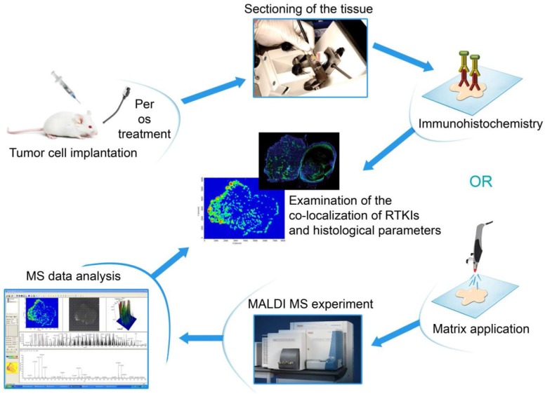
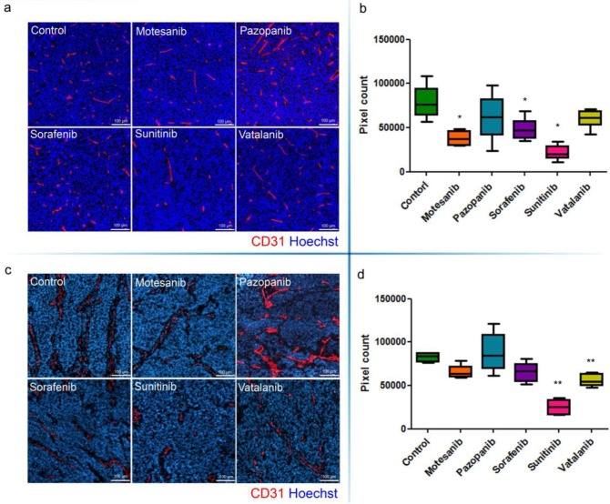
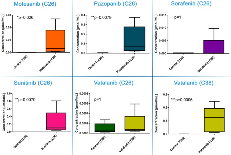
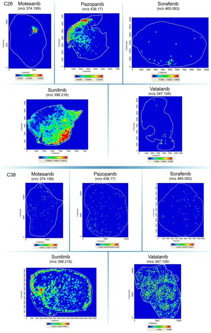
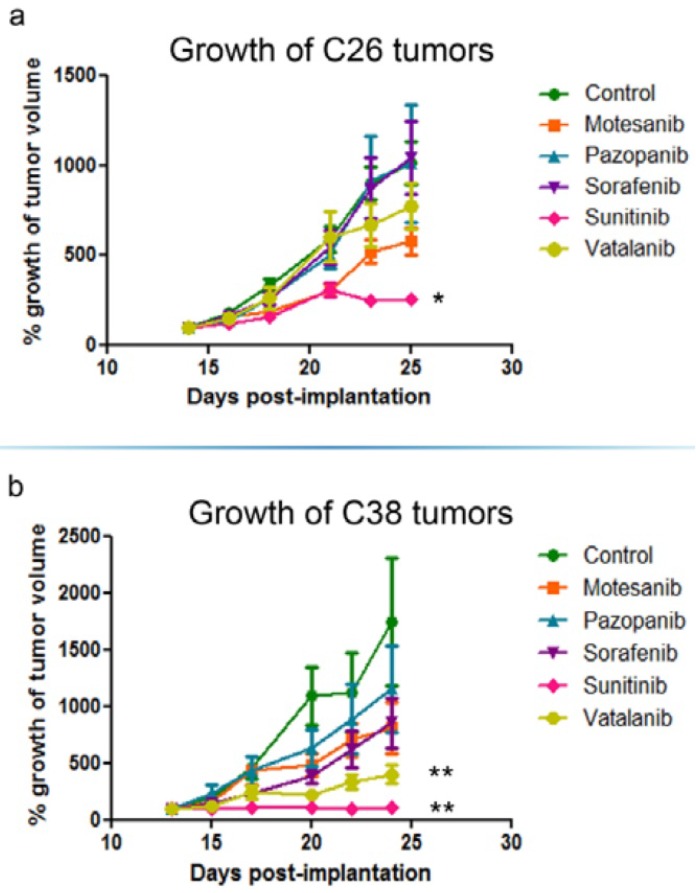
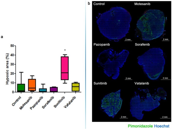
Resistance mechanisms against antiangiogenic drugs are unclear. Here, we correlated the antitumor and antivascular properties of five different antiangiogenic receptor tyrosine kinase inhibitors (RTKIs) (motesanib, pazopanib, sorafenib, sunitinib, vatalanib) with their intratumoral distribution data obtained by matrix-assisted laser desorption ionization mass spectrometry imaging (MALDI-MSI). In the first mouse model, only sunitinib exhibited broad-spectrum antivascular and antitumor activities by simultaneously suppressing vascular endothelial growth factor receptor-2 (VEGFR2) and desmin expression, and by increasing intratumoral hypoxia and inhibiting both tumor growth and vascularisation significantly. Importantly, the highest and most homogeneous intratumoral drug concentrations have been found in sunitinib-treated animals. In another animal model, where - in contrast to the first model - vatalanib was detectable at homogeneously high intratumoral concentrations, the drug significantly reduced tumor growth and angiogenesis. In conclusion, the tumor tissue penetration and thus the antiangiogenic and antitumor potential of antiangiogenic RTKIs vary among the tumor models and our study demonstrates the potential of MALDI-MSI to predict the efficacy of unlabelled small molecule antiangiogenic drugs in malignant tissue. Our approach is thus a major technical and preclinical advance demonstrating that primary resistance to angiogenesis inhibitors involves limited tumor tissue drug penetration. We also conclude that MALDI-MSI may significantly contribute to the improvement of antivascular cancer therapies.
抗血管生成药物的耐药机制尚不清楚。在此,我们将五种不同的抗血管生成受体酪氨酸激酶抑制剂(RTKIs)(莫替沙尼、帕唑帕尼、索拉非尼、舒尼替尼、凡德他尼)的抗肿瘤和抗血管特性与其通过基质辅助激光解吸电离质谱成像(MALDI-MSI)获得的瘤内分布数据进行了关联。在第一个小鼠模型中,只有舒尼替尼通过同时抑制血管内皮生长因子受体-2(VEGFR2)和结蛋白表达,以及增加瘤内缺氧并显著抑制肿瘤生长和血管生成,表现出广谱抗血管和抗肿瘤活性。重要的是,在接受舒尼替尼治疗的动物中发现了最高且最均匀的瘤内药物浓度。在另一个动物模型中,与第一个模型不同,凡德他尼在瘤内浓度均匀较高的情况下可检测到,该药物显著降低了肿瘤生长和血管生成。总之,抗血管生成RTKIs的肿瘤组织穿透率以及因此产生的抗血管生成和抗肿瘤潜力在不同肿瘤模型中有所不同,我们的研究证明了MALDI-MSI预测未标记小分子抗血管生成药物在恶性组织中疗效的潜力。因此,我们的方法是一项重大的技术和临床前进展,表明对血管生成抑制剂的原发性耐药涉及肿瘤组织药物穿透有限。我们还得出结论,MALDI-MSI可能会显著有助于改进抗血管生成癌症治疗。