Absalan Forouzan, Saremy Sadegh, Mansori Esrafil, Taheri Moghadam Mahin, Eftekhari Moghadam Ali Reza, Ghanavati Razie
Department of Anatomical Science, Faculty of Medicine, Ahvaz Jundishapur University of Medical Sciences, Ahvaz, Iran; Cellular and Molecular Research Center, Faculty of Medicine, Ahvaz Jundishapur University of Medical Sciences, Ahvaz, Iran.
Cellular and Molecular Research Center, Faculty of Medicine, Ahvaz Jundishapur University of Medical Sciences, Ahvaz, Iran.
Cell J. 2017 Winter;18(4):503-513. doi: 10.22074/cellj.2016.4717. Epub 2016 Sep 26.
Phthalates, which are commonly used to render plastics into soft and flexible materials, have also been determined as developmental and reproductive toxicants in human and animals. The purpose of this study was to evaluate the effect of mono-(2- ethylhexyl) phthalate (MEHP) and di-(2-ethylhexyl) phthalate (DEHP) oral administrations on maturation of mouse oocytes, apoptosis and gene transcription levels.
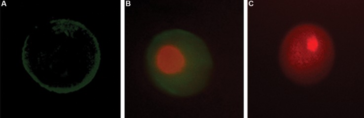
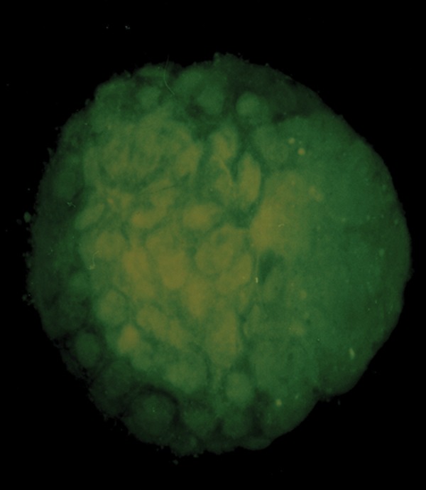
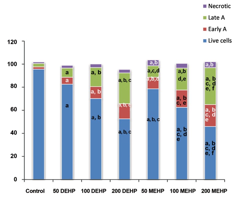
In this experimental study, immature oocytes recovered from Naval Medical Research Institute (NMRI) mouse strain (6-8 weeks), were divided into seven different experimental and control groups. Control group oocytes were retrieved from mice that received only normal saline. The experimental groups I, II or III oocytes were retrieved from mice treated with 50, 100 or 200 µl DEHP (2.56 µM) solution, respectively. The experimental groups IV, V or VI oocytes were retrieved from mouse exposed to 50, 100 or 200 µl MEHP (2.56 µM) solution, respectively. Fertilization and embryonic development were carried out in OMM and T6 medium. Apoptosis was assessed by annexin V-FITC/Dead Cell Apoptosis Kit, with PI staining. In addition, the mRNA levels of and were examined in oocytes. Finally, mouse embryo at early blastocyst stage was stained with acridine-orange (AO) and ethidium-bromide (EB), in order to access their viability.
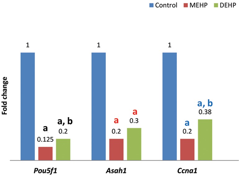
The proportion of oocytes that progressed up to metaphase II (MII) and 2-cells embryo formation stage was significantly decreased by exposure to MEHP or DEHP, in a dose-dependent manner. Annexin V and PI positive oocytes showed greater quantity in the treated mice than control. Quantitative reverse transcriptase-polymerase chain reaction (qRT-PCR) revealed that expression levels of and were significantly lower in the treated mouse oocytes than control. The total cell count for blastocyst developed from the treated mouse oocytes was lower than the controls.
These results indicate that oral administration of MEHP and DEHP could negatively affect mouse oocyte meiotic maturation and development , suggesting that phthalates could be risk factors for mammalians' reproductive health. Additionally, phthalate-induced changes in and transcription level could explain in part, the reduced developmental ability of mouse-treated oocytes.
邻苯二甲酸盐常用于使塑料变成柔软且有弹性的材料,它也被确定为人类和动物的发育及生殖毒物。本研究的目的是评估口服单(2-乙基己基)邻苯二甲酸酯(MEHP)和二(2-乙基己基)邻苯二甲酸酯(DEHP)对小鼠卵母细胞成熟、凋亡及基因转录水平的影响。
在本实验研究中,从海军医学研究所(NMRI)小鼠品系(6-8周龄)获取的未成熟卵母细胞被分为七个不同的实验组和对照组。对照组卵母细胞取自仅接受生理盐水的小鼠。实验组I、II或III的卵母细胞分别取自用50、100或200微升DEHP(2.56微摩尔)溶液处理的小鼠。实验组IV、V或VI的卵母细胞分别取自暴露于50、100或200微升MEHP(2.56微摩尔)溶液的小鼠。受精和胚胎发育在OMM和T6培养基中进行。通过膜联蛋白V-FITC/死细胞凋亡试剂盒及PI染色评估凋亡情况。此外,检测了卵母细胞中相关基因的mRNA水平。最后,用吖啶橙(AO)和溴化乙锭(EB)对早期囊胚阶段的小鼠胚胎进行染色,以评估其活力。
暴露于MEHP或DEHP后,发育至中期II(MII)和2-细胞胚胎形成阶段的卵母细胞比例以剂量依赖方式显著降低。处理组小鼠中膜联蛋白V和PI阳性的卵母细胞数量多于对照组。定量逆转录聚合酶链反应(qRT-PCR)显示,处理组小鼠卵母细胞中相关基因的表达水平显著低于对照组。处理组小鼠卵母细胞发育而来的囊胚总细胞数低于对照组。
这些结果表明,口服MEHP和DEHP会对小鼠卵母细胞减数分裂成熟和发育产生负面影响,提示邻苯二甲酸盐可能是哺乳动物生殖健康的危险因素。此外,邻苯二甲酸盐诱导的相关基因转录水平变化可能部分解释了处理组小鼠卵母细胞发育能力降低的原因。