• 文献检索
  • 文档翻译
  • 深度研究
  • 学术资讯
  • Suppr Zotero 插件Zotero 插件
  • 邀请有礼
  • 套餐&价格
  • 历史记录
应用&插件
Suppr Zotero 插件Zotero 插件浏览器插件Mac 客户端Windows 客户端微信小程序
定价
高级版会员购买积分包购买API积分包
服务
文献检索文档翻译深度研究API 文档MCP 服务
关于我们
关于 Suppr公司介绍联系我们用户协议隐私条款
关注我们

Suppr 超能文献

核心技术专利:CN118964589B侵权必究
粤ICP备2023148730 号-1Suppr @ 2026

文献检索

告别复杂PubMed语法,用中文像聊天一样搜索,搜遍4000万医学文献。AI智能推荐,让科研检索更轻松。

立即免费搜索

文件翻译

保留排版,准确专业,支持PDF/Word/PPT等文件格式,支持 12+语言互译。

免费翻译文档

深度研究

AI帮你快速写综述,25分钟生成高质量综述,智能提取关键信息,辅助科研写作。

立即免费体验

徒手B模式和功率模式三维超声用于可视化和评估颈内动脉狭窄的研究

Evaluation of Freehand B-Mode and Power-Mode 3D Ultrasound for Visualisation and Grading of Internal Carotid Artery Stenosis.

作者信息

Pelz Johann Otto, Weinreich Anna, Karlas Thomas, Saur Dorothee

机构信息

Department of Neurology, Leipzig University Hospital, Leipzig, Germany.

Department of Gastroenterology and Rheumatology, Leipzig University Hospital, Leipzig, Germany.

出版信息

PLoS One. 2017 Jan 3;12(1):e0167500. doi: 10.1371/journal.pone.0167500. eCollection 2017.

DOI:10.1371/journal.pone.0167500
PMID:28045903
原文链接:https://pmc.ncbi.nlm.nih.gov/articles/PMC5207436/
Abstract

BACKGROUND

Currently, colour-coded duplex sonography (2D-CDS) is clinical standard for detection and grading of internal carotid artery stenosis (ICAS). However, unlike angiographic imaging modalities, 2D-CDS assesses ICAS by its hemodynamic effects rather than luminal changes. Aim of this study was to evaluate freehand 3D ultrasound (3DUS) for direct visualisation and quantification of ICAS.

METHODS

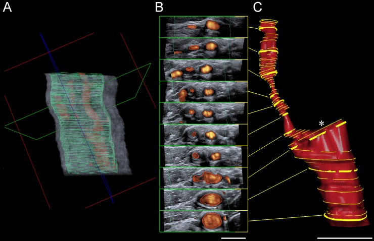
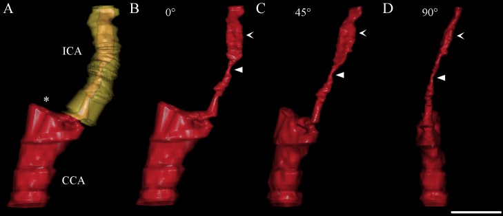
Thirty-seven patients with 43 ICAS were examined with 2D-CDS as reference standard and with freehand B-mode respectively power-mode 3DUS. Stenotic value of 3D reconstructed ICAS was calculated as distal diameter respectively distal cross-sectional area (CSA) reduction percentage and compared with 2D-CDS.

RESULTS

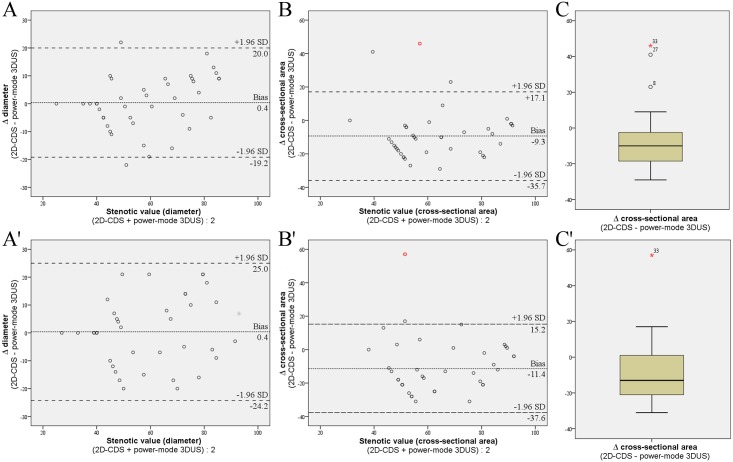
There was a trend but no significant difference in successful 3D reconstruction of ICAS between B-mode and power mode (examiner 1 {Ex1} 81% versus 93%, examiner 2 {Ex2} 84% versus 88%). Inter-rater agreement was best for power-mode 3DUS and assessment of stenotic value as distal CSA reduction percentage (intraclass correlation coefficient {ICC} 0.90) followed by power-mode 3DUS and distal diameter reduction percentage (ICC 0.81). Inter-rater agreement was poor for B-mode 3DUS (ICC, distal CSA reduction 0.36, distal diameter reduction 0.51). Intra-rater agreement for power-mode 3DUS was good for both measuring methods (ICC, distal CSA reduction 0.88 {Ex1} and 0.78 {Ex2}; ICC, distal diameter reduction 0.83 {Ex1} and 0.76 {Ex2}). In comparison to 2D-CDS inter-method agreement was good and clearly better for power-mode 3DUS (ICC, distal diameter reduction percentage: Ex1 0.85, Ex2 0.78; distal CSA reduction percentage: Ex1 0.63, Ex2 0.57) than for B-mode 3DUS (ICC, distal diameter reduction percentage: Ex1 0.40, Ex2 0.52; distal CSA reduction percentage: Ex1 0.15, Ex2 0.51).

CONCLUSIONS

Non-invasive power-mode 3DUS is superior to B-mode 3DUS for imaging and quantification of ICAS. Thereby, further studies are warranted which should now compare power-mode 3DUS with the angiographic gold standard imaging modalities for quantification of ICAS, i.e. with CTA or CE-MRA.

摘要

背景

目前,彩色编码双功超声检查(2D-CDS)是检测和分级颈内动脉狭窄(ICAS)的临床标准。然而,与血管造影成像方式不同,2D-CDS通过其血流动力学效应而非管腔变化来评估ICAS。本研究的目的是评估徒手三维超声(3DUS)用于直接可视化和量化ICAS。

方法

以2D-CDS作为参考标准,对37例患有43处ICAS的患者分别进行徒手B模式或能量模式3DUS检查。3D重建ICAS的狭窄值计算为远端直径或远端横截面积(CSA)减少百分比,并与2D-CDS进行比较。

结果

B模式和能量模式在ICAS的成功3D重建方面有一定趋势,但无显著差异(检查者1{Ex1}为81%对93%,检查者2{Ex2}为84%对88%)。评分者间一致性在能量模式3DUS以及将狭窄值评估为远端CSA减少百分比时最佳(组内相关系数{ICC}为0.90),其次是能量模式3DUS和远端直径减少百分比(ICC为0.81)。B模式3DUS的评分者间一致性较差(ICC,远端CSA减少为0.36,远端直径减少为0.51)。能量模式3DUS的评分者内一致性在两种测量方法中均良好(ICC,远端CSA减少:Ex1为0.88,Ex2为0.78;ICC,远端直径减少:Ex1为0.83,Ex2为0.76)。与2D-CDS相比, 方法间一致性良好,能量模式三维超声明显优于B模式三维超声(ICC,远端直径减少百分比:Ex1为0.85,Ex2为0.78;远端CSA减少百分比:Ex1为0.63,Ex2为0.57),而B模式三维超声(ICC,远端直径减少百分比:Ex1为0.40,Ex2为0.52;远端CSA减少百分比:Ex1为0.15,Ex2为0.51)。

结论

无创能量模式3DUS在ICAS成像和量化方面优于B模式3DUS。因此,有必要进行进一步研究,现在应将能量模式3DUS与用于ICAS量化的血管造影金标准成像方式,即CTA或CE-MRA进行比较。

https://cdn.ncbi.nlm.nih.gov/pmc/blobs/0799/5207436/afe1ac836d15/pone.0167500.g003.jpg
https://cdn.ncbi.nlm.nih.gov/pmc/blobs/0799/5207436/3c5ebf57da50/pone.0167500.g001.jpg
https://cdn.ncbi.nlm.nih.gov/pmc/blobs/0799/5207436/79601436ce4f/pone.0167500.g002.jpg
https://cdn.ncbi.nlm.nih.gov/pmc/blobs/0799/5207436/afe1ac836d15/pone.0167500.g003.jpg
https://cdn.ncbi.nlm.nih.gov/pmc/blobs/0799/5207436/3c5ebf57da50/pone.0167500.g001.jpg
https://cdn.ncbi.nlm.nih.gov/pmc/blobs/0799/5207436/79601436ce4f/pone.0167500.g002.jpg
https://cdn.ncbi.nlm.nih.gov/pmc/blobs/0799/5207436/afe1ac836d15/pone.0167500.g003.jpg

相似文献

1
Evaluation of Freehand B-Mode and Power-Mode 3D Ultrasound for Visualisation and Grading of Internal Carotid Artery Stenosis.徒手B模式和功率模式三维超声用于可视化和评估颈内动脉狭窄的研究
PLoS One. 2017 Jan 3;12(1):e0167500. doi: 10.1371/journal.pone.0167500. eCollection 2017.
2
Quantification of Internal Carotid Artery Stenosis with 3D Ultrasound Angiography.三维超声血管造影术对颈内动脉狭窄的定量分析
Ultraschall Med. 2015 Oct;36(5):487-93. doi: 10.1055/s-0034-1398749. Epub 2015 Jan 21.
3
Evaluation of the interrater and intermethod agreement of the German multiparametric ultrasound criteria for the grading of internal carotid artery stenosis.评价德国多参数超声标准评估颈内动脉狭窄程度的评级的评价者间和方法间一致性。
Neuroradiology. 2021 Apr;63(4):519-528. doi: 10.1007/s00234-020-02546-1. Epub 2020 Sep 18.
4
Multiparametric 3D Contrast-Enhanced Ultrasound to Assess Internal Carotid Artery Stenosis: A Pilot Study.应用三维对比增强超声多参数评估颈内动脉狭窄:一项初步研究。
J Neuroimaging. 2020 Jan;30(1):82-89. doi: 10.1111/jon.12662. Epub 2019 Sep 9.
5
Three-dimensional assessment of extracranial Doppler sonography in carotid artery stenosis compared with digital subtraction angiography.与数字减影血管造影术相比,颈动脉狭窄的颅外多普勒超声三维评估
Stroke. 2004 Aug;35(8):1847-51. doi: 10.1161/01.STR.0000133248.71808.38. Epub 2004 Jun 17.
6
High-grade stenoses of the internal carotid artery: comparison of high-resolution contrast enhanced 3D MRA, duplex sonography and power Doppler imaging.颈内动脉高度狭窄:高分辨率对比增强三维磁共振血管造影、双功超声及能量多普勒成像的比较
Eur J Radiol. 2006 Dec;60(3):379-86. doi: 10.1016/j.ejrad.2006.07.012. Epub 2006 Aug 21.
7
Neck Vessel Cross-Sectional Area Measured with MRI: Scan-Rescan Reproducibility for Longitudinal Evaluations.通过MRI测量的颈部血管横截面积:纵向评估的扫描-再扫描可重复性
J Neuroimaging. 2018 Jan;28(1):48-56. doi: 10.1111/jon.12488. Epub 2017 Dec 4.
8
CTA quantification of internal carotid artery stenosis: application of luminal area vs. luminal diameter measurements and assessment of inter-observer variability.颈内动脉狭窄的CT血管造影定量分析:管腔面积与管腔直径测量的应用及观察者间变异性评估
J Neuroimaging. 2007 Jul;17(3):219-26. doi: 10.1111/j.1552-6569.2007.00124.x.
9
Grading internal carotid artery stenosis using B-mode ultrasound (in vivo study).使用B型超声评估颈内动脉狭窄(体内研究)。
Eur J Vasc Endovasc Surg. 1999 Oct;18(4):315-22. doi: 10.1053/ejvs.1999.0884.
10
Assessing internal carotid artery stenosis with a semiautomated computed tomography angiography tool and duplex ultrasound.使用半自动计算机断层扫描血管造影工具和双功超声评估颈内动脉狭窄。
J Vasc Surg. 2015 Jun;61(6):1449-56. doi: 10.1016/j.jvs.2015.01.028. Epub 2015 Mar 14.

引用本文的文献

1
3D Ultrasound and MRI in Assessing Resection Margins during Tongue Cancer Surgery: A Research Protocol for a Clinical Diagnostic Accuracy Study.3D超声和MRI在舌癌手术中评估切缘的应用:一项临床诊断准确性研究的研究方案
J Imaging. 2023 Aug 28;9(9):174. doi: 10.3390/jimaging9090174.
2
Recent Advances in Tracking Devices for Biomedical Ultrasound Imaging Applications.用于生物医学超声成像应用的跟踪设备的最新进展。
Micromachines (Basel). 2022 Oct 29;13(11):1855. doi: 10.3390/mi13111855.
3
Spatiotemporal reconstruction method of carotid artery ultrasound from freehand sonography.

本文引用的文献

1
25 Years of 3D ultrasound in prenatal diagnosis (1989-2014).三维超声在产前诊断中的25年(1989 - 2014年)
Ultraschall Med. 2015 Feb;36(1):3-8. doi: 10.1055/s-0034-1398866. Epub 2015 Feb 5.
2
Quantification of Internal Carotid Artery Stenosis with 3D Ultrasound Angiography.三维超声血管造影术对颈内动脉狭窄的定量分析
Ultraschall Med. 2015 Oct;36(5):487-93. doi: 10.1055/s-0034-1398749. Epub 2015 Jan 21.
3
Global, regional, and national age-sex specific all-cause and cause-specific mortality for 240 causes of death, 1990-2013: a systematic analysis for the Global Burden of Disease Study 2013.
基于自由手超声的颈动脉超声时空重建方法。
Int J Comput Assist Radiol Surg. 2022 Sep;17(9):1731-1743. doi: 10.1007/s11548-022-02672-6. Epub 2022 Jun 15.
4
Feasibility of evaluating the stenosis of intracranial segment in the vertebral artery using neck-brain integrated ultrasound: a comparison with computed tomography angiography and digital subtraction angiography.颈部-脑部一体化超声评估椎动脉颅内段狭窄的可行性:与计算机断层扫描血管造影和数字减影血管造影的比较
Ann Transl Med. 2020 Sep;8(18):1155. doi: 10.21037/atm-20-5713.
5
Automated 3D geometry segmentation of the healthy and diseased carotid artery in free-hand, probe tracked ultrasound images.徒手、探头跟踪超声图像中健康和病变颈动脉的自动 3D 几何分割。
Med Phys. 2020 Mar;47(3):1034-1047. doi: 10.1002/mp.13960. Epub 2020 Jan 3.
6
Evaluation of a novel tomographic ultrasound device for abdominal examinations.新型断层超声成像设备在腹部检查中的评估。
PLoS One. 2019 Jun 26;14(6):e0218754. doi: 10.1371/journal.pone.0218754. eCollection 2019.
全球、地区和国家按年龄、性别划分的 240 种死因的全死因和特定死因死亡率,1990-2013 年:2013 年全球疾病负担研究的系统分析。
Lancet. 2015 Jan 10;385(9963):117-71. doi: 10.1016/S0140-6736(14)61682-2. Epub 2014 Dec 18.
4
Global and regional burden of stroke during 1990-2010: findings from the Global Burden of Disease Study 2010.1990-2010 年全球及各区域卒中负担变化:来自 2010 年全球疾病负担研究的结果。
Lancet. 2014 Jan 18;383(9913):245-54. doi: 10.1016/s0140-6736(13)61953-4.
5
Status update and interim results from the asymptomatic carotid surgery trial-2 (ACST-2).无症状性颈动脉手术试验-2(ACST-2)的现状更新和中期结果。
Eur J Vasc Endovasc Surg. 2013 Nov;46(5):510-8. doi: 10.1016/j.ejvs.2013.07.020. Epub 2013 Sep 17.
6
Guidelines for the early management of patients with acute ischemic stroke: a guideline for healthcare professionals from the American Heart Association/American Stroke Association.急性缺血性脑卒中患者早期管理指南:美国心脏协会/美国卒中协会医疗保健专业人员指南。
Stroke. 2013 Mar;44(3):870-947. doi: 10.1161/STR.0b013e318284056a. Epub 2013 Jan 31.
7
Three-dimensional ultrasound scanning.三维超声扫描。
Interface Focus. 2011 Aug 6;1(4):503-19. doi: 10.1098/rsfs.2011.0019. Epub 2011 Jun 1.
8
Grading carotid stenosis using ultrasonic methods.使用超声方法对颈动脉狭窄进行分级。
Stroke. 2012 Mar;43(3):916-21. doi: 10.1161/STROKEAHA.111.636084. Epub 2012 Feb 16.
9
Advantages and pitfalls of three-dimensional ultrasound imaging of carotid bifurcation.颈动脉分叉三维超声成像的优缺点。
Eur Neurol. 2011;65(6):309-16. doi: 10.1159/000327694. Epub 2011 May 17.
10
[Revision of DEGUM ultrasound criteria for grading internal carotid artery stenoses and transfer to NASCET measurement].[颈内动脉狭窄分级的DEGUM超声标准修订及向NASCET测量方法的转换]
Ultraschall Med. 2010 Jun;31(3):251-7. doi: 10.1055/s-0029-1245336. Epub 2010 Apr 22.