Gavidia-Bovadilla Giovana, Kanaan-Izquierdo Samir, Mataró-Serrat María, Perera-Lluna Alexandre
Department of ESAII, Universitat Politècnica de Catalunya, Barcelona, Catalonia, Spain.
Department of ESAII, Center for Biomedical Engineering Research (CREB), Universitat Politècnica de Catalunya, Barcelona, Catalonia, Spain.
PLoS One. 2017 Jan 3;12(1):e0168011. doi: 10.1371/journal.pone.0168011. eCollection 2017.
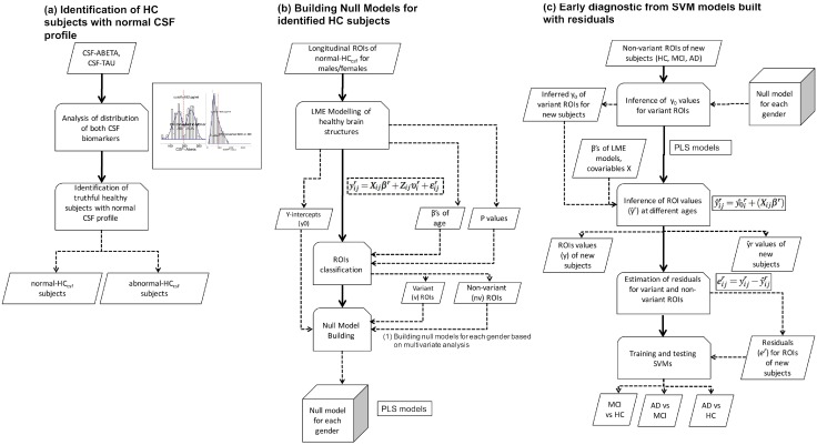
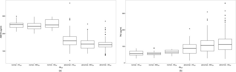
Incipient Alzheimer's Disease (AD) is characterized by a slow onset of clinical symptoms, with pathological brain changes starting several years earlier. Consequently, it is necessary to first understand and differentiate age-related changes in brain regions in the absence of disease, and then to support early and accurate AD diagnosis. However, there is poor understanding of the initial stage of AD; seemingly healthy elderly brains lose matter in regions related to AD, but similar changes can also be found in non-demented subjects having mild cognitive impairment (MCI). By using a Linear Mixed Effects approach, we modelled the change of 166 Magnetic Resonance Imaging (MRI)-based biomarkers available at a 5-year follow up on healthy elderly control (HC, n = 46) subjects. We hypothesized that, by identifying their significant variant (vr) and quasi-variant (qvr) brain regions over time, it would be possible to obtain an age-based null model, which would characterize their normal atrophy and growth patterns as well as the correlation between these two regions. By using the null model on those subjects who had been clinically diagnosed as HC (n = 161), MCI (n = 209) and AD (n = 331), normal age-related changes were estimated and deviation scores (residuals) from the observed MRI-based biomarkers were computed. Subject classification, as well as the early prediction of conversion to MCI and AD, were addressed through residual-based Support Vector Machines (SVM) modelling. We found reductions in most cortical volumes and thicknesses (with evident gender differences) as well as in sub-cortical regions, including greater atrophy in the hippocampus. The average accuracies (ACC) recorded for men and women were: AD-HC: 94.11%, MCI-HC: 83.77% and MCI converted to AD (cAD)-MCI non-converter (sMCI): 76.72%. Likewise, as compared to standard clinical diagnosis methods, SVM classifiers predicted the conversion of cAD to be 1.9 years earlier for females (ACC:72.5%) and 1.4 years earlier for males (ACC:69.0%).
早期阿尔茨海默病(AD)的临床症状起病缓慢,而大脑的病理变化在数年前就已开始。因此,有必要首先了解和区分无疾病情况下大脑区域与年龄相关的变化,进而支持AD的早期准确诊断。然而,人们对AD的初始阶段了解甚少;看似健康的老年大脑在与AD相关的区域会出现物质流失,但在患有轻度认知障碍(MCI)的非痴呆受试者中也能发现类似变化。通过使用线性混合效应方法,我们对健康老年对照(HC,n = 46)受试者在5年随访中可获得的166种基于磁共振成像(MRI)的生物标志物的变化进行了建模。我们假设,通过识别随时间变化的显著变异(vr)和准变异(qvr)脑区,有可能获得一个基于年龄的零模型,该模型将表征其正常萎缩和生长模式以及这两个区域之间的相关性。通过对临床诊断为HC(n = 161)、MCI(n = 209)和AD(n = 331)的受试者使用零模型,估计了正常的年龄相关变化,并计算了基于观察到的MRI生物标志物的偏差分数(残差)。通过基于残差的支持向量机(SVM)建模解决了受试者分类以及向MCI和AD转化的早期预测问题。我们发现大多数皮质体积和厚度(存在明显的性别差异)以及皮质下区域均减少,包括海马体萎缩更明显。男性和女性记录的平均准确率(ACC)分别为:AD - HC:94.11%,MCI - HC:83.77%,MCI转化为AD(cAD) - MCI未转化者(sMCI):76.72%。同样,与标准临床诊断方法相比,SVM分类器预测女性cAD转化提前1.9年(ACC:72.5%),男性提前1.4年(ACC:69.0%)。