Silva Bruno Jorge de Andrade, Barbosa Mayara Garcia de Mattos, Andrade Priscila Ribeiro, Ferreira Helen, Nery José Augusto da Costa, Côrte-Real Suzana, da Silva Gilberto Marcelo Sperandio, Rosa Patricia Sammarco, Fabri Mario, Sarno Euzenir Nunes, Pinheiro Roberta Olmo
Leprosy Laboratory; Oswaldo Cruz Institute; Oswaldo Cruz Foundation, FIOCRUZ; Rio de Janeiro, Brazil.
Structural Biology Laboratory; Oswaldo Cruz Institute; Oswaldo Cruz Foundation, FIOCRUZ; Rio de Janeiro, Brazil.
PLoS Pathog. 2017 Jan 5;13(1):e1006103. doi: 10.1371/journal.ppat.1006103. eCollection 2017 Jan.
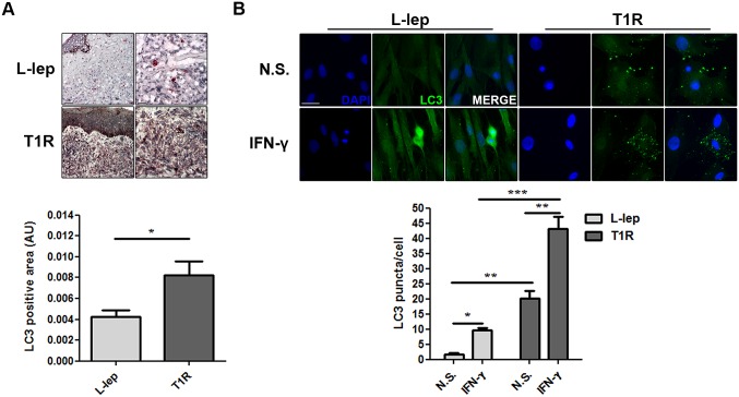
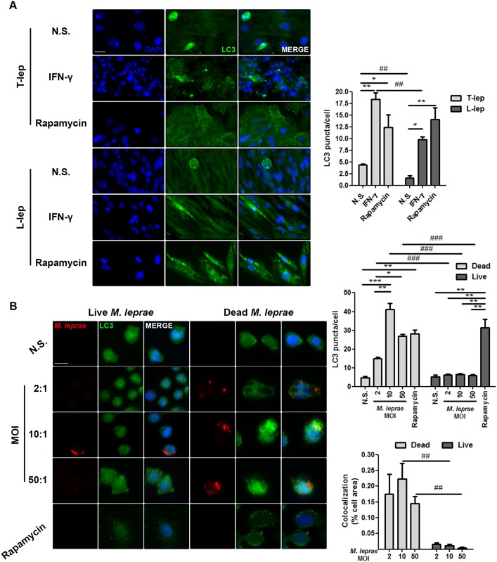
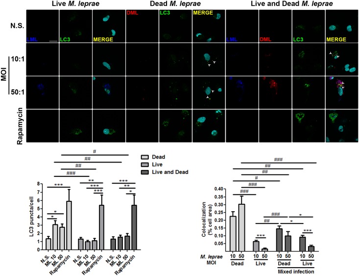
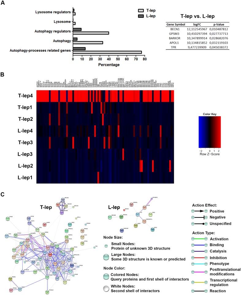
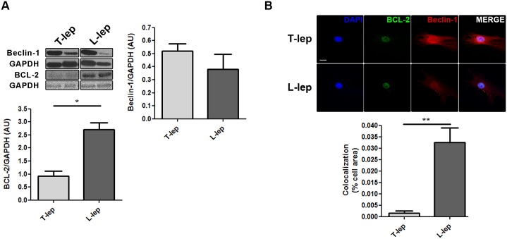
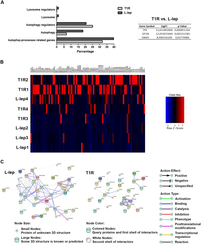
Leprosy is a chronic infectious disease that may present different clinical forms according to the immune response of the host. Levels of IFN-γ are significantly raised in paucibacillary tuberculoid (T-lep) when compared with multibacillary lepromatous (L-lep) patients. IFN-γ primes macrophages for inflammatory activation and induces the autophagy antimicrobial mechanism. The involvement of autophagy in the immune response against Mycobacterium leprae remains unexplored. Here, we demonstrated by different autophagic assays that LC3-positive autophagosomes were predominantly observed in T-lep when compared with L-lep lesions and skin-derived macrophages. Accumulation of the autophagic receptors SQSTM1/p62 and NBR1, expression of lysosomal antimicrobial peptides and colocalization analysis of autolysosomes revealed an impairment of the autophagic flux in L-lep cells, which was restored by IFN-γ or rapamycin treatment. Autophagy PCR array gene-expression analysis revealed a significantly upregulation of autophagy genes (BECN1, GPSM3, ATG14, APOL1, and TPR) in T-lep cells. Furthermore, an upregulation of autophagy genes (TPR, GFI1B and GNAI3) as well as LC3 levels was observed in cells of L-lep patients that developed type 1 reaction (T1R) episodes, an acute inflammatory condition associated with increased IFN-γ levels. Finally, we observed increased BCL2 expression in L-lep cells that could be responsible for the blockage of BECN1-mediated autophagy. In addition, in vitro studies demonstrated that dead, but not live M. leprae can induce autophagy in primary and lineage human monocytes, and that live mycobacteria can reduce the autophagy activation triggered by dead mycobacteria, suggesting that M. leprae may hamper the autophagic machinery as an immune escape mechanism. Together, these results indicate that autophagy is an important innate mechanism associated with the M. leprae control in skin macrophages.
麻风是一种慢性传染病,根据宿主的免疫反应可能呈现不同的临床形式。与多菌型瘤型(L-麻风)患者相比,少菌型结核样型(T-麻风)患者的γ干扰素水平显著升高。γ干扰素使巨噬细胞启动炎症激活并诱导自噬抗菌机制。自噬在抗麻风分枝杆菌免疫反应中的作用仍未得到充分研究。在此,我们通过不同的自噬检测方法证明,与L-麻风病变和皮肤来源的巨噬细胞相比,T-麻风病变中主要观察到LC3阳性自噬体。自噬受体SQSTM1/p62和NBR1的积累、溶酶体抗菌肽的表达以及自噬溶酶体的共定位分析显示L-麻风细胞中自噬流受损,而γ干扰素或雷帕霉素处理可恢复这种受损情况。自噬PCR阵列基因表达分析显示T-麻风细胞中自噬基因(BECN1、GPSM3、ATG14、APOL1和TPR)显著上调。此外,在发生1型反应(T1R)发作(一种与γ干扰素水平升高相关的急性炎症状态)的L-麻风患者细胞中,观察到自噬基因(TPR、GFI1B和GNAI3)以及LC3水平上调。最后,我们观察到L-麻风细胞中BCL2表达增加,这可能是导致BECN1介导的自噬受阻的原因。此外,体外研究表明,死的而非活的麻风分枝杆菌可诱导原代和谱系人单核细胞发生自噬,并且活的分枝杆菌可降低死的分枝杆菌触发的自噬激活,这表明麻风分枝杆菌可能作为一种免疫逃逸机制阻碍自噬机制。总之,这些结果表明自噬是皮肤巨噬细胞中与控制麻风分枝杆菌相关的重要固有机制。