• 文献检索
  • 文档翻译
  • 深度研究
  • 学术资讯
  • Suppr Zotero 插件Zotero 插件
  • 邀请有礼
  • 套餐&价格
  • 历史记录
应用&插件
Suppr Zotero 插件Zotero 插件浏览器插件Mac 客户端Windows 客户端微信小程序
定价
高级版会员购买积分包购买API积分包
服务
文献检索文档翻译深度研究API 文档MCP 服务
关于我们
关于 Suppr公司介绍联系我们用户协议隐私条款
关注我们

Suppr 超能文献

核心技术专利:CN118964589B侵权必究
粤ICP备2023148730 号-1Suppr @ 2026

文献检索

告别复杂PubMed语法,用中文像聊天一样搜索,搜遍4000万医学文献。AI智能推荐,让科研检索更轻松。

立即免费搜索

文件翻译

保留排版,准确专业,支持PDF/Word/PPT等文件格式,支持 12+语言互译。

免费翻译文档

深度研究

AI帮你快速写综述,25分钟生成高质量综述,智能提取关键信息,辅助科研写作。

立即免费体验

用安全无镉的 ZnS-AgInS 纳米粒子的水相 ZnS 涂层对移植的脂肪组织来源干细胞进行标记和体内可视化。

Labeling and in vivo visualization of transplanted adipose tissue-derived stem cells with safe cadmium-free aqueous ZnS coating of ZnS-AgInS nanoparticles.

机构信息

Department of Applied Chemistry, Graduate School of Engineering, Nagoya University, Furo-cho, Chikusa-ku, Nagoya 464-8603, Japan.

ImPACT Research Center for Advanced Nanobiodevices, Nagoya University, Furo-cho, Chikusa-ku, Nagoya 464-8603, Japan.

出版信息

Sci Rep. 2017 Jan 6;7:40047. doi: 10.1038/srep40047.

DOI:10.1038/srep40047
PMID:28059135
原文链接:https://pmc.ncbi.nlm.nih.gov/articles/PMC5216330/
Abstract

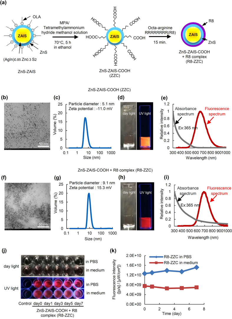
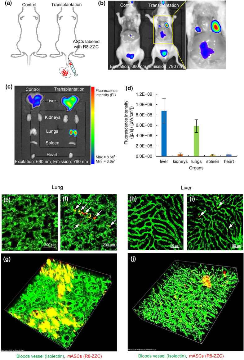
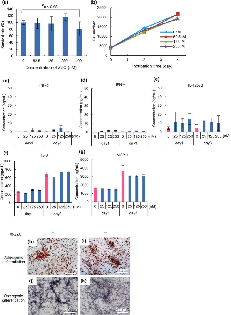
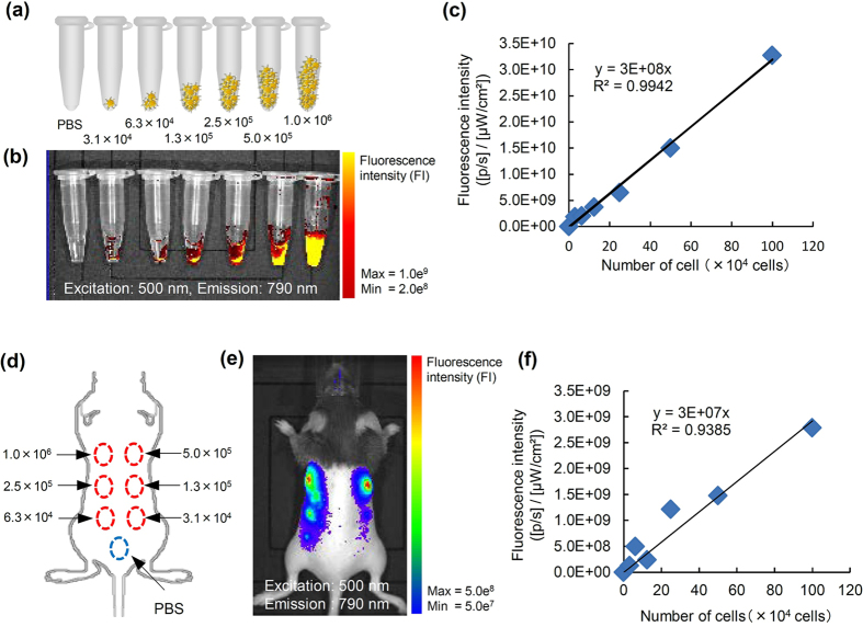
The facile synthesis of ZnS-AgInS (ZAIS) as cadmium-free QDs and their application, mainly in solar cells, has been reported by our groups. In the present study, we investigated the safety and the usefulness for labeling and in vivo imaging of a newly synthesized aqueous ZnS-coated ZAIS (ZnS-ZAIS) carboxylated nanoparticles (ZZC) to stem cells. ZZC shows the strong fluorescence in aqueous solutions such as PBS and cell culture medium, and a complex of ZZC and octa-arginine (R8) peptides (R8-ZZC) can achieve the highly efficient labeling of adipose tissue-derived stem cells (ASCs). The cytotoxicity of R8-ZZC to ASCs was found to be extremely low in comparison to that of CdSe-based QDs, and R8-ZZC was confirmed to have no influence on the proliferation rate or the differentiation ability of ASCs. Moreover, R8-ZZC was not found to induce the production of major inflammatory cytokines (TNF-α, IFN-γ, IL-12p70, IL-6 and MCP-1) in ASCs. Transplanted R8-ZZC-labeled ASCs could be quantitatively detected in the lungs and liver mainly using an in vivo imaging system. In addition, high-speed multiphoton confocal laser microscopy revealed the presence of aggregates of transplanted ASCs at many sites in the lungs, whereas individual ASCs were found to have accumulated in the liver.

摘要

我们小组已经报道了一种简便的方法来合成无镉的 ZnS-AgInS(ZAIS)量子点,并将其应用于太阳能电池等领域。在本研究中,我们研究了一种新合成的水溶性 ZnS 包覆的 ZAIS(ZnS-ZAIS)羧基化纳米粒子(ZZC)的安全性和在干细胞标记和体内成像方面的应用。ZZC 在 PBS 和细胞培养基等水溶液中具有很强的荧光,并且 ZZC 与八精氨酸(R8)肽(R8-ZZC)的复合物可以实现对脂肪组织来源干细胞(ASCs)的高效标记。与基于 CdSe 的量子点相比,R8-ZZC 对 ASCs 的细胞毒性极低,并且证实 R8-ZZC 对 ASCs 的增殖率和分化能力没有影响。此外,R8-ZZC 不会诱导 ASCs 产生主要的炎症细胞因子(TNF-α、IFN-γ、IL-12p70、IL-6 和 MCP-1)。使用体内成像系统主要可以定量检测移植到肺部和肝脏中的 R8-ZZC 标记的 ASCs。此外,高速多光子共聚焦激光显微镜显示,在肺部的许多部位都存在移植的 ASCs 聚集,而在肝脏中则发现单个 ASCs 聚集。

https://cdn.ncbi.nlm.nih.gov/pmc/blobs/4c49/5216330/37ac6fbcdf93/srep40047-f5.jpg
https://cdn.ncbi.nlm.nih.gov/pmc/blobs/4c49/5216330/12b53cce8b94/srep40047-f1.jpg
https://cdn.ncbi.nlm.nih.gov/pmc/blobs/4c49/5216330/e19194e3e9da/srep40047-f2.jpg
https://cdn.ncbi.nlm.nih.gov/pmc/blobs/4c49/5216330/0515c69be4c6/srep40047-f3.jpg
https://cdn.ncbi.nlm.nih.gov/pmc/blobs/4c49/5216330/1f3acae3d8bb/srep40047-f4.jpg
https://cdn.ncbi.nlm.nih.gov/pmc/blobs/4c49/5216330/37ac6fbcdf93/srep40047-f5.jpg

相似文献

1
Labeling and in vivo visualization of transplanted adipose tissue-derived stem cells with safe cadmium-free aqueous ZnS coating of ZnS-AgInS nanoparticles.用安全无镉的 ZnS-AgInS 纳米粒子的水相 ZnS 涂层对移植的脂肪组织来源干细胞进行标记和体内可视化。
Sci Rep. 2017 Jan 6;7:40047. doi: 10.1038/srep40047.
2
Adipose Tissue-Derived Stem Cell Imaging Using Cadmium-Free Quantum Dots.使用无镉量子点对脂肪组织来源的干细胞进行成像。
Cell Med. 2013 Oct 29;6(1-2):91-7. doi: 10.3727/215517913X674261. eCollection 2013 Dec 30.
3
Monitoring transplanted adipose tissue-derived stem cells combined with heparin in the liver by fluorescence imaging using quantum dots.荧光量子点成像监测肝内移植脂肪组织来源干细胞联合肝素。
Biomaterials. 2012 Mar;33(7):2177-86. doi: 10.1016/j.biomaterials.2011.12.009. Epub 2011 Dec 20.
4
Quantum dots labeling using octa-arginine peptides for imaging of adipose tissue-derived stem cells.使用八聚精氨酸肽对脂肪组织来源干细胞进行量子点标记成像。
Biomaterials. 2010 May;31(14):4094-103. doi: 10.1016/j.biomaterials.2010.01.134. Epub 2010 Feb 19.
5
Quantum dots for labeling adipose tissue-derived stem cells.用于标记脂肪组织来源干细胞的量子点
Cell Transplant. 2009;18(5):591-9. doi: 10.1177/096368970901805-615.
6
Novel positively charged nanoparticle labeling for in vivo imaging of adipose tissue-derived stem cells.用于脂肪组织来源干细胞体内成像的新型带正电荷纳米颗粒标记
PLoS One. 2014 Nov 3;9(11):e110142. doi: 10.1371/journal.pone.0110142. eCollection 2014.
7
Cadmium-free sugar-chain-immobilized fluorescent nanoparticles containing low-toxicity ZnS-AgInS2 cores for probing lectin and cells.无镉糖链固定化荧光纳米粒子,其内核含有低毒性的 ZnS-AgInS2,可用于探测凝集素和细胞。
Bioconjug Chem. 2014 Feb 19;25(2):286-95. doi: 10.1021/bc400425w. Epub 2014 Jan 30.
8
Transduction Function of a Magnetic Nanoparticle TMADM for Stem-Cell Imaging with Quantum Dots.用于量子点干细胞成像的磁性纳米颗粒TMADM的转导功能
Anal Sci. 2017;33(2):143-146. doi: 10.2116/analsci.33.143.
9
Estimation of the distribution of intravenously injected adipose tissue-derived stem cells labeled with quantum dots in mice organs through the determination of their metallic components by ICPMS.通过电感耦合等离子体质谱法(ICPMS)测定量子点标记的静脉内注射脂肪组织源性干细胞的金属成分来估计其在小鼠器官中的分布。
Anal Chem. 2011 Nov 1;83(21):8252-8. doi: 10.1021/ac202053y. Epub 2011 Oct 13.
10
Aqueous synthesis of highly luminescent AgInS₂-ZnS quantum dots and their biological applications.水相合成高发光的 AgInS₂-ZnS 量子点及其生物应用。
Nanoscale. 2013 Mar 21;5(6):2322-7. doi: 10.1039/c3nr34159c.

引用本文的文献

1
Establishment of a stem cell administration imaging method in bleomycin-induced pulmonary fibrosis mouse models.建立博来霉素诱导的肺纤维化小鼠模型中的干细胞给药成像方法。
Sci Rep. 2024 Aug 14;14(1):18905. doi: 10.1038/s41598-024-67586-6.
2
Transplantation of committed pre-adipocytes from brown adipose tissue improves whole-body glucose homeostasis.棕色脂肪组织中定向前脂肪细胞的移植可改善全身葡萄糖稳态。
iScience. 2024 Jan 17;27(2):108927. doi: 10.1016/j.isci.2024.108927. eCollection 2024 Feb 16.
3
From Red to Green Luminescence via Surface Functionalization. Effect of 2-(5-Mercaptothien-2-yl)-8-(thien-2-yl)-5-hexylthieno[3,4-]pyrrole-4,6-dione Ligands on the Photoluminescence of Alloyed Ag-In-Zn-S Nanocrystals.

本文引用的文献

1
In vivo biodistribution studies and ex vivo lymph node imaging using heavy metal-free quantum dots.使用无重金属量子点的体内生物分布研究和体外淋巴结成像。
Biomaterials. 2016 Oct;104:182-91. doi: 10.1016/j.biomaterials.2016.07.014. Epub 2016 Jul 12.
2
Synthesis of Hydrophilic CuInS2/ZnS Quantum Dots with Different Polymeric Shells and Study of Their Cytotoxicity and Hemocompatibility.具有不同聚合物壳层的亲水性CuInS2/ZnS量子点的合成及其细胞毒性和血液相容性研究。
ACS Appl Mater Interfaces. 2016 Mar;8(12):7613-22. doi: 10.1021/acsami.5b11258. Epub 2016 Mar 21.
3
Immunotoxicity assessment of CdSe/ZnS quantum dots in macrophages, lymphocytes and BALB/c mice.
通过表面功能化实现从红色到绿色发光。2-(5-巯基噻吩-2-基)-8-(噻吩-2-基)-5-己基噻吩并[3,4-]吡咯-4,6-二酮配体对合金化Ag-In-Zn-S纳米晶体光致发光的影响。
Inorg Chem. 2020 Oct 5;59(19):14594-14604. doi: 10.1021/acs.inorgchem.0c02468. Epub 2020 Sep 17.
4
The Composition-Dependent Photoluminescence Properties of Non-Stoichiometric ZnAgInS Nanocrystals.非化学计量比ZnAgInS纳米晶体的成分依赖光致发光特性
Micromachines (Basel). 2019 Jul 1;10(7):439. doi: 10.3390/mi10070439.
5
Click Chemistry as a Tool for Cell Engineering and Drug Delivery.点击化学作为细胞工程和药物传递的工具。
Molecules. 2019 Jan 4;24(1):172. doi: 10.3390/molecules24010172.
CdSe/ZnS量子点在巨噬细胞、淋巴细胞和BALB/c小鼠中的免疫毒性评估
J Nanobiotechnology. 2016 Feb 4;14:10. doi: 10.1186/s12951-016-0162-4.
4
Sunflower-type nanogels carrying a quantum dot nanoprobe for both superior gene delivery efficacy and tracing of human mesenchymal stem cells.葵花型纳米凝胶负载量子点纳米探针,兼具优异的基因转染效率和人骨髓间充质干细胞示踪效果。
Biomaterials. 2016 Jan;77:14-25. doi: 10.1016/j.biomaterials.2015.11.002. Epub 2015 Nov 2.
5
Carbon "Quantum" Dots for Fluorescence Labeling of Cells.用于细胞荧光标记的碳“量子”点
ACS Appl Mater Interfaces. 2015 Sep 2;7(34):19439-45. doi: 10.1021/acsami.5b05665. Epub 2015 Aug 18.
6
In vivo real-time visualization of mesenchymal stem cells tropism for cutaneous regeneration using NIR-II fluorescence imaging.利用近红外二区荧光成像技术在体实时可视化间充质干细胞对皮肤再生的趋向性。
Biomaterials. 2015 Jun;53:265-73. doi: 10.1016/j.biomaterials.2015.02.090. Epub 2015 Mar 16.
7
Quantum dot/antibody conjugates for in vivo cytometric imaging in mice.用于小鼠体内细胞计数成像的量子点/抗体缀合物。
Proc Natl Acad Sci U S A. 2015 Feb 3;112(5):1350-5. doi: 10.1073/pnas.1421632111. Epub 2015 Jan 20.
8
Facile synthesis of water-soluble Zn-doped AgIn5S8/ZnS core/shell fluorescent nanocrystals and their biological application.水溶性锌掺杂硫化银铟锌/硫化锌核壳荧光纳米晶体的简便合成及其生物学应用。
Inorg Chem. 2015 Feb 16;54(4):1627-33. doi: 10.1021/ic502600u. Epub 2015 Jan 16.
9
Migration mechanism of mesenchymal stem cells studied by QD/NSOM.
Biochim Biophys Acta. 2015 Mar;1848(3):859-68. doi: 10.1016/j.bbamem.2014.12.013. Epub 2014 Dec 19.
10
Precise and long-term tracking of adipose-derived stem cells and their regenerative capacity via superb bright and stable organic nanodots.通过超亮稳定的有机纳米点精确且长期追踪脂肪来源干细胞及其再生能力。
ACS Nano. 2014 Dec 23;8(12):12620-31. doi: 10.1021/nn505554y. Epub 2014 Nov 26.