Zhang Qian, Waqas Yasir, Yang Ping, Sun Xuejing, Liu Yi, Ahmed Nisar, Chen Bing, Li Quanfu, Hu Lisi, Huang Yufei, Chen Hong, Hu Bing, Chen Qiusheng
Laboratory of Animal Cell Biology and Embryology, College of Veterinary Medicine, Nanjing Agricultural University, Nanjing, China.
Key Laboratory of Antibody Techniques of Ministry of Health, Nanjing Medical University, Nanjing, China.
Oncotarget. 2017 Jan 31;8(5):7405-7419. doi: 10.18632/oncotarget.14502.
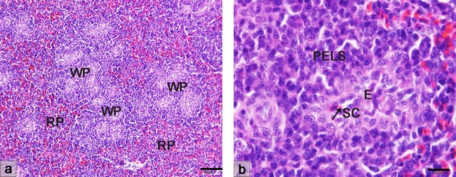
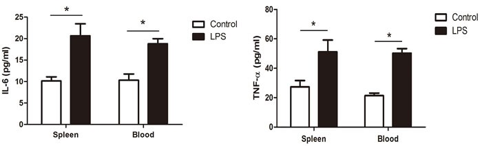
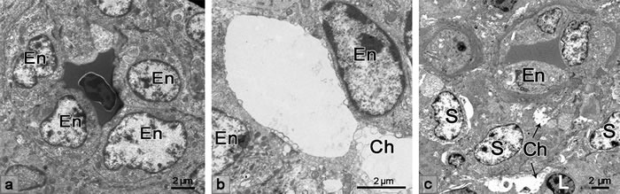
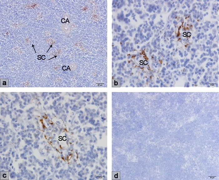
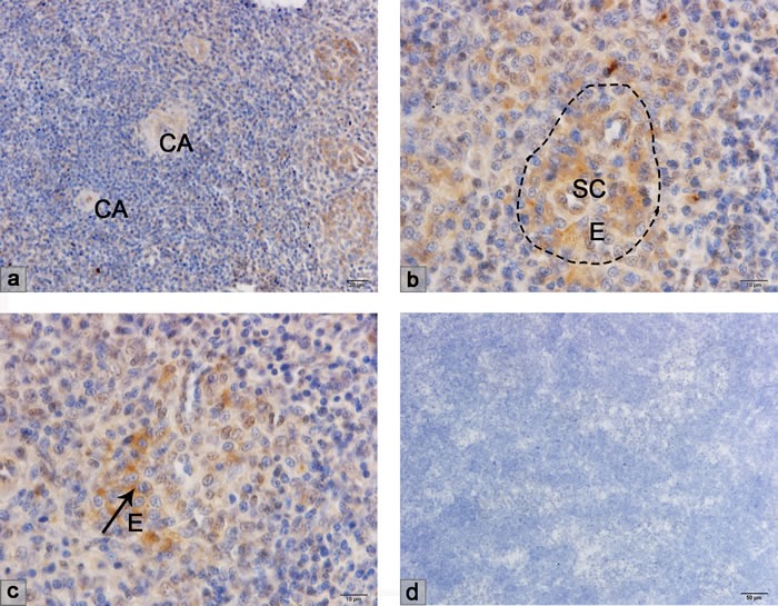
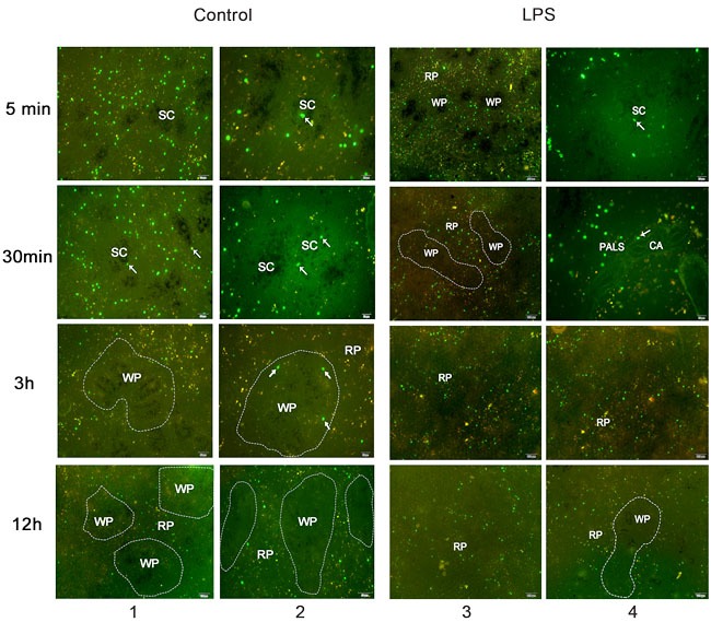
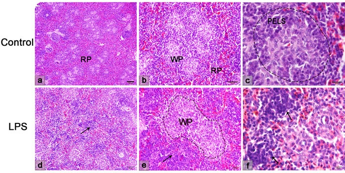
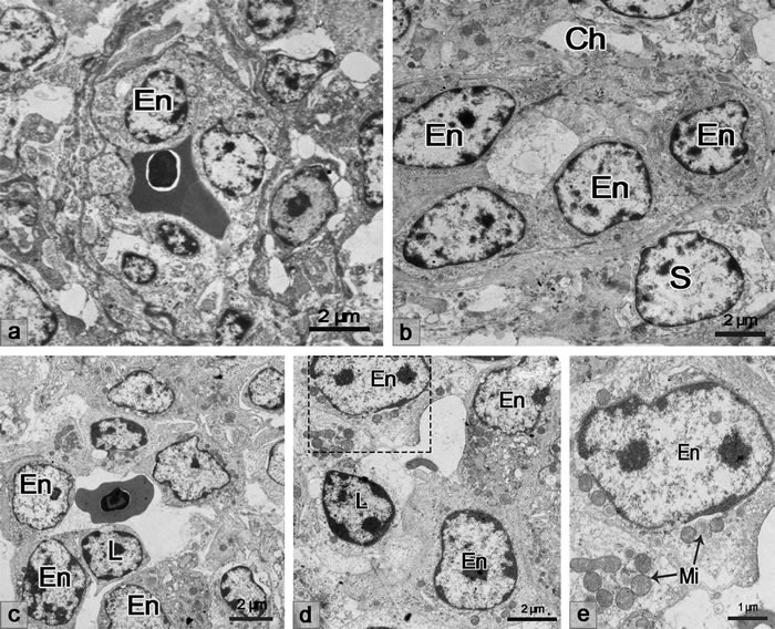
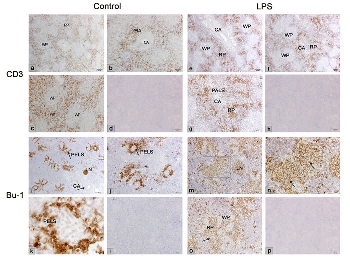
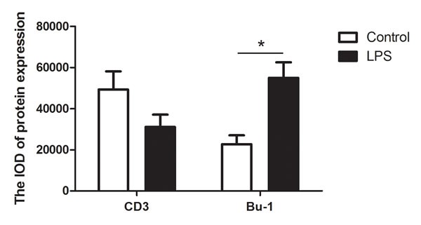
The immune function of the chicken spleen depends on its different compartments of red and white pulps, but little is known about the mechanism underlying lymphocyte homing towards the different compartments. In the present study, the role of lymphocyte homing in the chicken spleen was investigated during lipopolysaccharide (LPS) stimulation. Morphological analysis demonstrated the cuboidal endothelial cells of the splenic sheathed capillary facilitated the passage of lymphocyte homing to the chicken spleen. The tissue-specific adhesion molecules- vascular cell adhesion molecule-1 (VCAM-1) and mucosal addressin cell adhesion molecule-1 (MADCAM-1) expressed on the sheathed capillary, which suggested the high endothelial venule (HEV)-like vessels of the chicken spleen. Electron microscope analysis showed LPS activated the endothelium of the sheathed capillary and recruited lymphocytes to the chicken spleen. Transferring of 5, 6- carboxyfluorescein diacetate, succinimidyl ester (CFSE) labeled lymphocytes depicted the rout of lymphocyte homing to the compartments of the chicken spleen was from the white pulp to the red pulp. Furthermore, the mRNA and protein levels of adhesion molecular integrin β1 and VCAM-1 increased after LPS stimulation. The mechanism underlying the integrin β1 and VCAM-1 during LPS stimulation might be associated with the integrin linked kinase (ILK)- dependent regulation of protein kinase B (PKB/AKT). This study firstly shows lymphocyte homing in the chicken spleen after LPS-induced inflammation. These results contribute to our knowledge of comparative immunology and provide a better means for investigating the pharmacological strategies concerning the possible role of lymphocyte homing in inflammation and immunological reactions in infectious disease.
鸡脾脏的免疫功能取决于其红髓和白髓的不同区域,但关于淋巴细胞归巢至不同区域的潜在机制知之甚少。在本研究中,我们调查了脂多糖(LPS)刺激期间淋巴细胞归巢在鸡脾脏中的作用。形态学分析表明,脾被膜毛细血管的立方内皮细胞促进了淋巴细胞归巢至鸡脾脏的过程。在被膜毛细血管上表达的组织特异性黏附分子——血管细胞黏附分子-1(VCAM-1)和黏膜地址素细胞黏附分子-1(MADCAM-1),提示鸡脾脏存在类似高内皮微静脉(HEV)的血管。电子显微镜分析显示,LPS激活了被膜毛细血管的内皮,并将淋巴细胞募集至鸡脾脏。5,6-羧基荧光素二乙酸琥珀酰亚胺酯(CFSE)标记淋巴细胞的转移显示,淋巴细胞归巢至鸡脾脏各区域的途径是从白髓到红髓。此外,LPS刺激后黏附分子整合素β1和VCAM-1的mRNA和蛋白水平升高。LPS刺激期间整合素β1和VCAM-1的潜在机制可能与整合素连接激酶(ILK)依赖的蛋白激酶B(PKB/AKT)调节有关。本研究首次展示了LPS诱导炎症后鸡脾脏中的淋巴细胞归巢。这些结果有助于我们了解比较免疫学,并为研究淋巴细胞归巢在传染病炎症和免疫反应中可能作用的药理策略提供了更好的手段。