• 文献检索
  • 文档翻译
  • 深度研究
  • 学术资讯
  • Suppr Zotero 插件Zotero 插件
  • 邀请有礼
  • 套餐&价格
  • 历史记录
应用&插件
Suppr Zotero 插件Zotero 插件浏览器插件Mac 客户端Windows 客户端微信小程序
定价
高级版会员购买积分包购买API积分包
服务
文献检索文档翻译深度研究API 文档MCP 服务
关于我们
关于 Suppr公司介绍联系我们用户协议隐私条款
关注我们

Suppr 超能文献

核心技术专利:CN118964589B侵权必究
粤ICP备2023148730 号-1Suppr @ 2026

文献检索

告别复杂PubMed语法,用中文像聊天一样搜索,搜遍4000万医学文献。AI智能推荐,让科研检索更轻松。

立即免费搜索

文件翻译

保留排版,准确专业,支持PDF/Word/PPT等文件格式,支持 12+语言互译。

免费翻译文档

深度研究

AI帮你快速写综述,25分钟生成高质量综述,智能提取关键信息,辅助科研写作。

立即免费体验

使用多分辨率染色颜色表示的统计分析进行染色反卷积

Stain Deconvolution Using Statistical Analysis of Multi-Resolution Stain Colour Representation.

作者信息

Alsubaie Najah, Trahearn Nicholas, Raza Shan E Ahmed, Snead David, Rajpoot Nasir M

机构信息

Department of Computer Science, University of Warwick, Coventry, United Kingdom.

Department of Computer Science, Princess Nourah University, Riyadh, Kingdom of Saudi Arabia.

出版信息

PLoS One. 2017 Jan 11;12(1):e0169875. doi: 10.1371/journal.pone.0169875. eCollection 2017.

DOI:10.1371/journal.pone.0169875
PMID:28076381
原文链接:https://pmc.ncbi.nlm.nih.gov/articles/PMC5226799/
Abstract

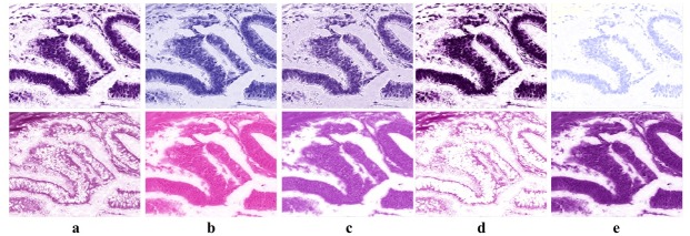
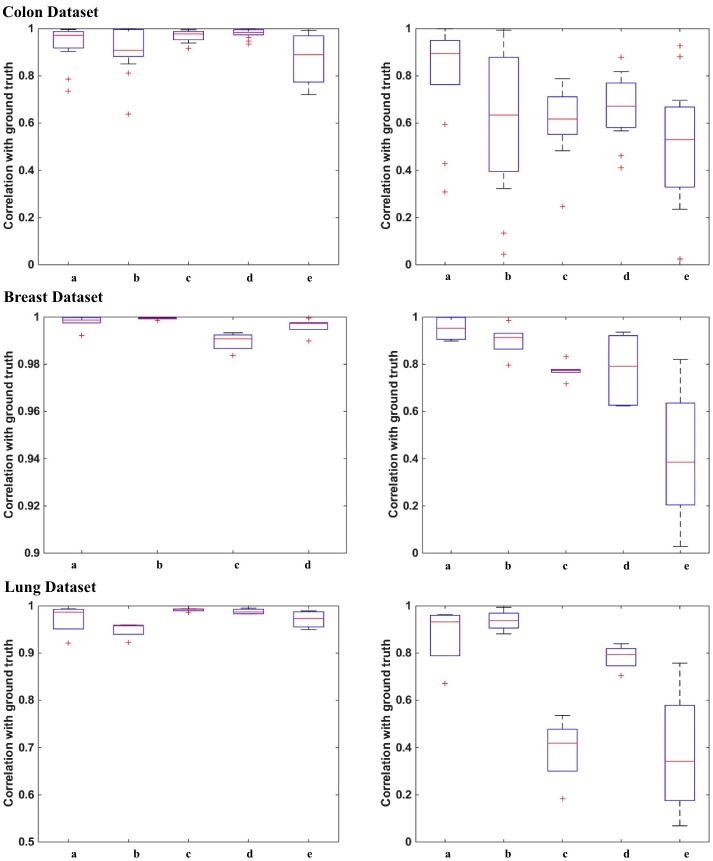
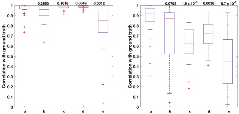
Stain colour estimation is a prominent factor of the analysis pipeline in most of histology image processing algorithms. Providing a reliable and efficient stain colour deconvolution approach is fundamental for robust algorithm. In this paper, we propose a novel method for stain colour deconvolution of histology images. This approach statistically analyses the multi-resolutional representation of the image to separate the independent observations out of the correlated ones. We then estimate the stain mixing matrix using filtered uncorrelated data. We conducted an extensive set of experiments to compare the proposed method to the recent state of the art methods and demonstrate the robustness of this approach using three different datasets of scanned slides, prepared in different labs using different scanners.

摘要

染色颜色估计是大多数组织学图像处理算法中分析流程的一个重要因素。提供一种可靠且高效的染色颜色反卷积方法对于稳健的算法至关重要。在本文中,我们提出了一种用于组织学图像染色颜色反卷积的新方法。该方法对图像的多分辨率表示进行统计分析,以从相关观测值中分离出独立观测值。然后,我们使用滤波后的不相关数据估计染色混合矩阵。我们进行了大量实验,将所提出的方法与最新的现有方法进行比较,并使用在不同实验室使用不同扫描仪制备的三个不同扫描玻片数据集证明了该方法的稳健性。

https://cdn.ncbi.nlm.nih.gov/pmc/blobs/9400/5226799/f364f5d37889/pone.0169875.g007.jpg
https://cdn.ncbi.nlm.nih.gov/pmc/blobs/9400/5226799/89db24d00793/pone.0169875.g001.jpg
https://cdn.ncbi.nlm.nih.gov/pmc/blobs/9400/5226799/dec5e4102397/pone.0169875.g002.jpg
https://cdn.ncbi.nlm.nih.gov/pmc/blobs/9400/5226799/04137438acf3/pone.0169875.g003.jpg
https://cdn.ncbi.nlm.nih.gov/pmc/blobs/9400/5226799/472ddceb6ef4/pone.0169875.g004.jpg
https://cdn.ncbi.nlm.nih.gov/pmc/blobs/9400/5226799/8e80bb4c5652/pone.0169875.g005.jpg
https://cdn.ncbi.nlm.nih.gov/pmc/blobs/9400/5226799/9e645b23163e/pone.0169875.g006.jpg
https://cdn.ncbi.nlm.nih.gov/pmc/blobs/9400/5226799/f364f5d37889/pone.0169875.g007.jpg

相似文献

1
Stain Deconvolution Using Statistical Analysis of Multi-Resolution Stain Colour Representation.使用多分辨率染色颜色表示的统计分析进行染色反卷积
PLoS One. 2017 Jan 11;12(1):e0169875. doi: 10.1371/journal.pone.0169875. eCollection 2017.
2
Normalization of HE-stained histological images using cycle consistent generative adversarial networks.使用循环一致生成对抗网络对 HE 染色组织学图像进行归一化。
Diagn Pathol. 2021 Aug 6;16(1):71. doi: 10.1186/s13000-021-01126-y.
3
Unsupervised method for normalization of hematoxylin-eosin stain in histological images.苏木精-伊红染色组织图像的无监督归一化方法。
Comput Med Imaging Graph. 2019 Oct;77:101646. doi: 10.1016/j.compmedimag.2019.101646. Epub 2019 Aug 14.
4
A nonlinear mapping approach to stain normalization in digital histopathology images using image-specific color deconvolution.一种使用特定图像颜色反卷积对数字组织病理学图像进行染色归一化的非线性映射方法。
IEEE Trans Biomed Eng. 2014 Jun;61(6):1729-38. doi: 10.1109/TBME.2014.2303294.
5
Appearance normalization of histology slides.组织学切片的外观标准化
Comput Med Imaging Graph. 2015 Jul;43:89-98. doi: 10.1016/j.compmedimag.2015.03.005. Epub 2015 Mar 21.
6
Breast histopathology image segmentation using spatio-colour-texture based graph partition method.基于空间颜色纹理的图划分方法的乳腺组织病理学图像分割
J Microsc. 2016 Jun;262(3):260-73. doi: 10.1111/jmi.12361. Epub 2015 Dec 28.
7
Bayesian K-SVD for H and E blind color deconvolution. Applications to stain normalization, data augmentation and cancer classification.贝叶斯 K-SVD 用于 H 和 E 盲色反卷积。在染色归一化、数据增强和癌症分类中的应用。
Comput Med Imaging Graph. 2022 Apr;97:102048. doi: 10.1016/j.compmedimag.2022.102048. Epub 2022 Feb 15.
8
Variational Bayesian Blind Color Deconvolution of Histopathological Images.变分贝叶斯盲彩色组织病理学图像反卷积。
IEEE Trans Image Process. 2020;29(1):2026-2036. doi: 10.1109/TIP.2019.2946442. Epub 2019 Oct 15.
9
Colour adaptive generative networks for stain normalisation of histopathology images.用于组织病理学图像染色归一化的颜色自适应生成网络。
Med Image Anal. 2022 Nov;82:102580. doi: 10.1016/j.media.2022.102580. Epub 2022 Aug 27.
10
Truncated Normal Mixture Prior Based Deep Latent Model for Color Normalization of Histology Images.基于截断正态混合先验的组织学图像颜色归一化深度潜在模型。
IEEE Trans Med Imaging. 2023 Jun;42(6):1746-1757. doi: 10.1109/TMI.2023.3238425. Epub 2023 Jun 1.

引用本文的文献

1
Unsupervised Stain Decomposition via Inversion Regulation for Multiplex Immunohistochemistry Images.通过反演调节实现的多重免疫组织化学图像无监督染色分解
Proc Mach Learn Res. 2023 Jul;227:74-94.
2
Oncogenic ETS fusions promote DNA damage and proinflammatory responses via pericentromeric RNAs in extracellular vesicles.致癌性 ETS 融合通过细胞外囊泡中的着丝粒周围 RNA 促进 DNA 损伤和促炎反应。
J Clin Invest. 2024 Mar 26;134(9):e169470. doi: 10.1172/JCI169470.
3
A Robust Method for the Unsupervised Scoring of Immunohistochemical Staining.

本文引用的文献

1
Locality Sensitive Deep Learning for Detection and Classification of Nuclei in Routine Colon Cancer Histology Images.基于局部敏感的深度学习在常规结肠癌组织学图像中细胞核检测与分类的应用。
IEEE Trans Med Imaging. 2016 May;35(5):1196-1206. doi: 10.1109/TMI.2016.2525803. Epub 2016 Feb 4.
2
New Colors for Histology: Optimized Bivariate Color Maps Increase Perceptual Contrast in Histological Images.组织学的新色彩:优化的双变量色图增强组织学图像中的感知对比度
PLoS One. 2015 Dec 30;10(12):e0145572. doi: 10.1371/journal.pone.0145572. eCollection 2015.
3
Blind colour separation of H&E stained histological images by linearly transforming the colour space.
一种用于免疫组织化学染色无监督评分的稳健方法。
Entropy (Basel). 2024 Feb 15;26(2):165. doi: 10.3390/e26020165.
4
Towards Artificial Intelligence Applications in Next Generation Cytopathology.迈向人工智能在下一代细胞病理学中的应用
Biomedicines. 2023 Aug 8;11(8):2225. doi: 10.3390/biomedicines11082225.
5
Enhanced Pathology Image Quality with Restore-Generative Adversarial Network.基于修复生成对抗网络的增强病理学图像质量
Am J Pathol. 2023 Apr;193(4):404-416. doi: 10.1016/j.ajpath.2022.12.011. Epub 2023 Jan 18.
6
Deep learning-based framework for slide-based histopathological image analysis.基于深度学习的幻灯片组织病理学图像分析框架。
Sci Rep. 2022 Nov 9;12(1):19075. doi: 10.1038/s41598-022-23166-0.
7
Breast histopathological image analysis using image processing techniques for diagnostic puposes: A methodological review.基于图像处理技术的乳腺组织病理图像分析在诊断中的应用:方法学综述。
J Med Syst. 2021 Dec 3;46(1):7. doi: 10.1007/s10916-021-01786-9.
8
Quantitative and Semi-quantitative Methods for Assessing the Degree of Methylene Blue Staining in Sentinel Lymph Nodes in Dogs.评估犬前哨淋巴结亚甲蓝染色程度的定量和半定量方法
Front Vet Sci. 2021 Oct 21;8:758295. doi: 10.3389/fvets.2021.758295. eCollection 2021.
9
Automated annotations of epithelial cells and stroma in hematoxylin-eosin-stained whole-slide images using cytokeratin re-staining.使用细胞角蛋白重染对苏木精-伊红染色的全切片图像中的上皮细胞和基质进行自动注释。
J Pathol Clin Res. 2022 Mar;8(2):129-142. doi: 10.1002/cjp2.249. Epub 2021 Oct 30.
10
Deep Learning of Histopathology Images at the Single Cell Level.单细胞水平的组织病理学图像深度学习
Front Artif Intell. 2021 Sep 10;4:754641. doi: 10.3389/frai.2021.754641. eCollection 2021.
通过线性变换颜色空间对苏木精-伊红(H&E)染色的组织学图像进行盲色分离。
J Microsc. 2015 Dec;260(3):377-88. doi: 10.1111/jmi.12304. Epub 2015 Sep 10.
4
Stain Specific Standardization of Whole-Slide Histopathological Images.全切片组织病理学图像的染色特异性标准化
IEEE Trans Med Imaging. 2016 Feb;35(2):404-15. doi: 10.1109/TMI.2015.2476509. Epub 2015 Sep 4.
5
Appearance normalization of histology slides.组织学切片的外观标准化
Comput Med Imaging Graph. 2015 Jul;43:89-98. doi: 10.1016/j.compmedimag.2015.03.005. Epub 2015 Mar 21.
6
A Complete Color Normalization Approach to Histopathology Images Using Color Cues Computed From Saturation-Weighted Statistics.一种使用从饱和度加权统计计算出的颜色线索对组织病理学图像进行完全颜色归一化的方法。
IEEE Trans Biomed Eng. 2015 Jul;62(7):1862-73. doi: 10.1109/TBME.2015.2405791. Epub 2015 Feb 19.
7
Assessment of algorithms for mitosis detection in breast cancer histopathology images.乳腺癌组织病理学图像有丝分裂检测算法的评估。
Med Image Anal. 2015 Feb;20(1):237-48. doi: 10.1016/j.media.2014.11.010. Epub 2014 Nov 29.
8
A nonlinear mapping approach to stain normalization in digital histopathology images using image-specific color deconvolution.一种使用特定图像颜色反卷积对数字组织病理学图像进行染色归一化的非线性映射方法。
IEEE Trans Biomed Eng. 2014 Jun;61(6):1729-38. doi: 10.1109/TBME.2014.2303294.
9
Blind color decomposition of histological images.组织学图像的盲色彩分解。
IEEE Trans Med Imaging. 2013 Jun;32(6):983-94. doi: 10.1109/TMI.2013.2239655. Epub 2013 Jan 11.
10
Efficient nucleus detector in histopathology images.高效的组织病理学图像细胞核检测器。
J Microsc. 2013 Feb;249(2):124-35. doi: 10.1111/jmi.12001. Epub 2012 Dec 17.