Nelissen Jules L, de Graaf Larry, Traa Willeke A, Schreurs Tom J L, Moerman Kevin M, Nederveen Aart J, Sinkus Ralph, Oomens Cees W J, Nicolay Klaas, Strijkers Gustav J
Biomedical NMR, Biomedical Engineering, Eindhoven University of Technology, Eindhoven, The Netherlands.
Biomedical Engineering and Physics, Academic Medical Center, Amsterdam, The Netherlands.
PLoS One. 2017 Jan 11;12(1):e0169864. doi: 10.1371/journal.pone.0169864. eCollection 2017.
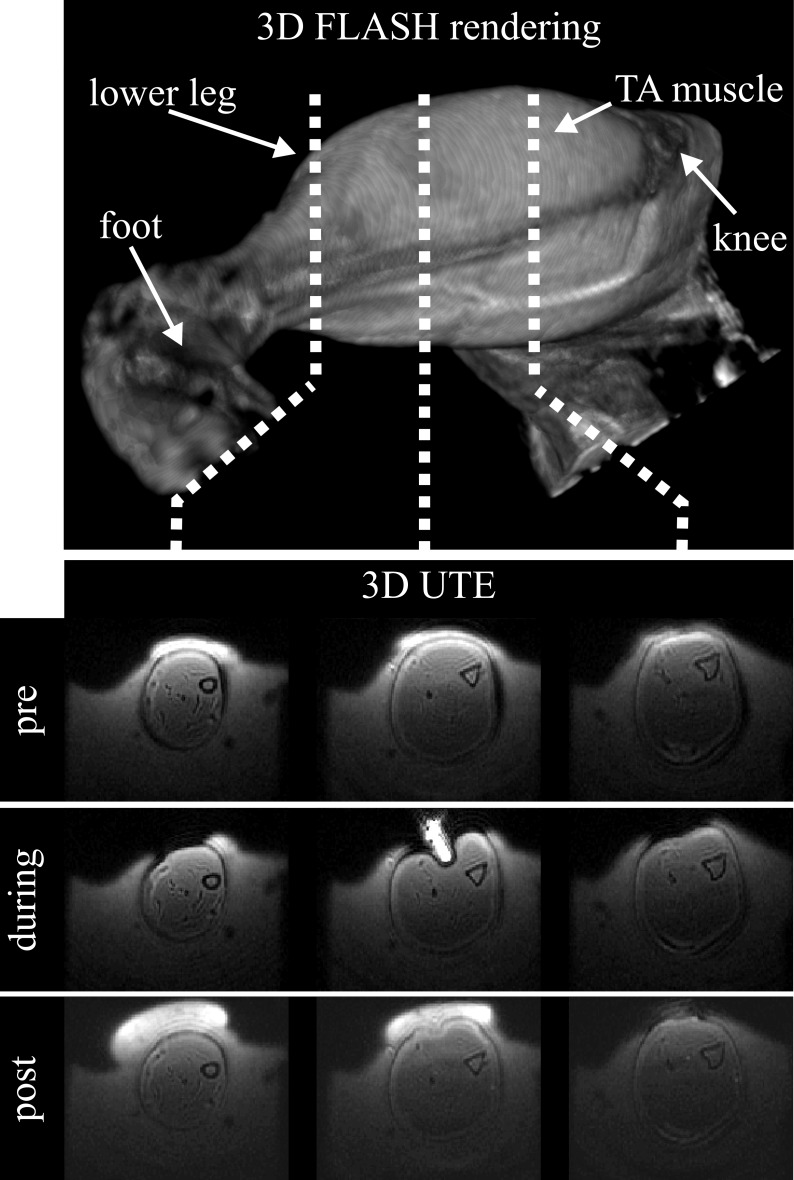
Deformation of skeletal muscle in the proximity of bony structures may lead to deep tissue injury category of pressure ulcers. Changes in mechanical properties have been proposed as a risk factor in the development of deep tissue injury and may be useful as a diagnostic tool for early detection. MRE allows for the estimation of mechanical properties of soft tissue through analysis of shear wave data. The shear waves originate from vibrations induced by an external actuator placed on the tissue surface. In this study a combined Magnetic Resonance (MR) compatible indentation and MR Elastography (MRE) setup is presented to study mechanical properties associated with deep tissue injury in rats. The proposed setup allows for MRE investigations combined with damage-inducing large strain indentation of the Tibialis Anterior muscle in the rat hind leg inside a small animal MR scanner. An alginate cast allowed proper fixation of the animal leg with anatomical perfect fit, provided boundary condition information for FEA and provided good susceptibility matching. MR Elastography data could be recorded for the Tibialis Anterior muscle prior to, during, and after indentation. A decaying shear wave with an average amplitude of approximately 2 μm propagated in the whole muscle. MRE elastograms representing local tissue shear storage modulus Gd showed significant increased mean values due to damage-inducing indentation (from 4.2 ± 0.1 kPa before to 5.1 ± 0.6 kPa after, p<0.05). The proposed setup enables controlled deformation under MRI-guidance, monitoring of the wound development by MRI, and quantification of tissue mechanical properties by MRE. We expect that improved knowledge of changes in soft tissue mechanical properties due to deep tissue injury, will provide new insights in the etiology of deep tissue injuries, skeletal muscle damage and other related muscle pathologies.
骨骼结构附近的骨骼肌变形可能导致压疮的深部组织损伤类别。机械性能的变化已被提出作为深部组织损伤发展的一个风险因素,并且可能作为早期检测的诊断工具。磁共振弹性成像(MRE)通过分析剪切波数据来估计软组织的机械性能。剪切波源于放置在组织表面的外部致动器引起的振动。在本研究中,提出了一种结合磁共振(MR)兼容压痕和磁共振弹性成像(MRE)的装置,以研究大鼠深部组织损伤相关的机械性能。所提出的装置允许在小动物磁共振扫描仪内对大鼠后腿的胫前肌进行MRE研究,并结合损伤诱导的大应变压痕。藻酸盐铸型能够以解剖学上的完美贴合正确固定动物腿部,提供有限元分析(FEA)的边界条件信息,并提供良好的磁化率匹配。在压痕之前、期间和之后都可以记录胫前肌的磁共振弹性成像数据。一个平均振幅约为2μm的衰减剪切波在整个肌肉中传播。代表局部组织剪切储存模量Gd的MRE弹性图显示,由于损伤诱导压痕,平均值显著增加(从之前的4.2±0.1kPa增加到之后的5.1±0.6kPa,p<0.05)。所提出的装置能够在MRI引导下进行可控变形,通过MRI监测伤口发展,并通过MRE对组织机械性能进行量化。我们期望,对深部组织损伤引起的软组织机械性能变化的深入了解,将为深部组织损伤、骨骼肌损伤和其他相关肌肉疾病的病因学提供新的见解。