Chen Shui-Wen, Fu Wei, Liu Jing, Wang Yan
aDepartment of Neonatology and NICU of Bayi Children's Hospital,the Army General Hospital of the Chinese PLA affiliated to Southern Medical University, Beijing bDepartment of Pediatrics, Shenzhen Baoan Maternal and Child Health Hospital, Shenzhen, China.
Medicine (Baltimore). 2017 Jan;96(2):e5826. doi: 10.1097/MD.0000000000005826.
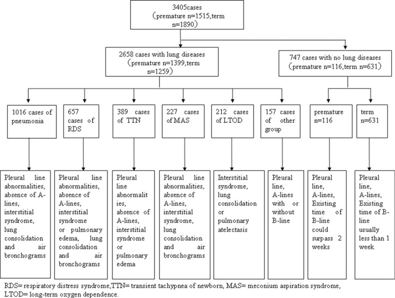
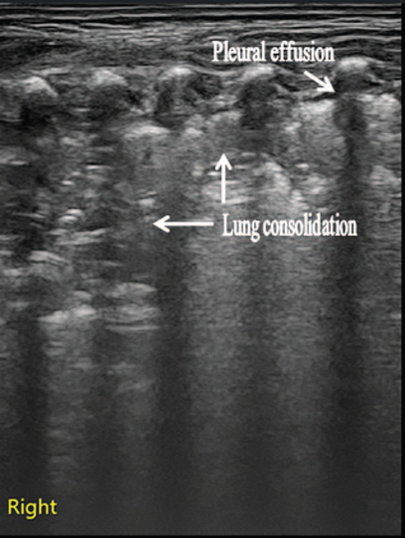
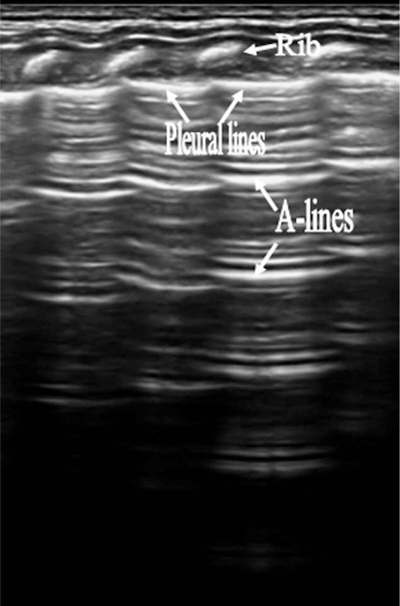
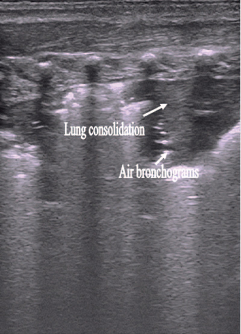
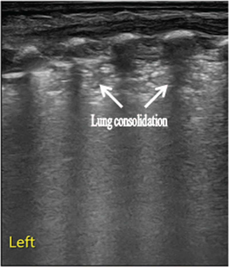
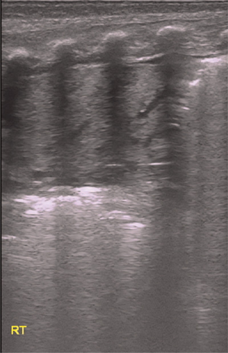
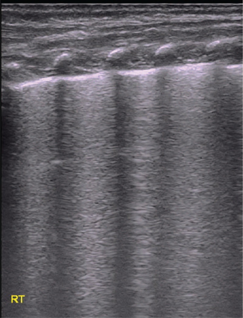
The aim of this study was to study the features of lung ultrasonography (LUS) in lung disease and to evaluate the usefulness of LUS in the neonatal intensive care unit (NICU).All of 3405 neonates included in this study underwent an LUS examination. Diagnoses were based on medical history, clinical manifestation, laboratory examination, and signs on chest radiography (CR) and/or computed tomography (CT). A single expert physician performed all LUS examinations.There were 2658 cases (78.9%) with lung disease and 747 cases (21.9%) without lung disease. The main signs of neonates with lung disease on LUS were as follows: absence of A-lines, pleural-line abnormalities, interstitial syndrome, lung consolidation, air bronchograms, pulmonary edema, and lung pulse. These abnormal signs were reduced or eliminated on LUS as patient conditions improved. There were 81 cases that could not be diagnosed as lung disease by CR but were discovered as pneumonia, respiratory distress syndrome (RDS), or transient tachypnea of newborn (TTN) on LUS. Likewise, 23 cases misdiagnosed as RDS by CR were diagnosed as TTN on LUS. Among 212 cases of long-term oxygen dependence (LTOD) that failed to yield signs of pulmonary edema and lung consolidation on CR, 103 cases showed abnormal signs on LUS. Among 747 cases without lung disease, B-lines of 713 neonates (95.4%) could be found within 3 days after birth, and 256 neonates (34.3%) could be observed from 3 days to 1 week after birth. B-lines of 19 cases could be detected from 1 to 2 weeks after birth. The longest time at which B-lines could still be observed was 19 days after birth.LUS has clinical value for the diagnosis of lung disease and the discrimination of causes of LTOP in premature infants, particularly for the diagnosis and identification of RDS and TTN. Moreover, LUS has additional advantages, including its lack of radiation exposure and its ability to noninvasively monitor treatment progress. Therefore, LUS should be routinely used in the NICU.
本研究旨在探讨肺部超声(LUS)在肺部疾病中的特征,并评估LUS在新生儿重症监护病房(NICU)的应用价值。本研究纳入的3405例新生儿均接受了LUS检查。诊断依据病史、临床表现、实验室检查以及胸部X线摄影(CR)和/或计算机断层扫描(CT)的征象。由一名专业医师进行所有LUS检查。有肺部疾病的病例2658例(78.9%),无肺部疾病的病例747例(21.9%)。LUS检查显示有肺部疾病的新生儿的主要征象如下:A线消失、胸膜线异常、间质综合征、肺实变、支气管充气征、肺水肿和肺搏动。随着病情改善,这些异常征象在LUS检查中减轻或消失。有81例经CR检查不能诊断为肺部疾病,但经LUS检查发现为肺炎、呼吸窘迫综合征(RDS)或新生儿短暂性呼吸急促(TTN)。同样,23例经CR误诊为RDS的病例经LUS检查诊断为TTN。在212例长期氧依赖(LTOD)病例中,CR检查未显示肺水肿和肺实变征象,但LUS检查有103例显示异常征象。在747例无肺部疾病的病例中,713例(95.4%)新生儿在出生后3天内可发现B线,256例(34.3%)在出生后3天至1周可观察到B线。19例在出生后1至2周可检测到B线。B线仍可观察到的最长时间为出生后19天。LUS对早产儿肺部疾病的诊断及长期氧依赖(LTOP)病因的鉴别具有临床价值,尤其对RDS和TTN的诊断及鉴别。此外,LUS还有其他优势,包括无辐射暴露以及能够无创监测治疗进展。因此,LUS应在NICU中常规使用。