Arthuis Chloé J, Novell Anthony, Raes Florian, Escoffre Jean-Michel, Lerondel Stéphanie, Le Pape Alain, Bouakaz Ayache, Perrotin Franck
Inserm U930, François Rabelais University, Tours, France.
University Hospital Center of Tours, Department of Obstetrics, Gynecology and Fetal Medicine, Tours, France.
PLoS One. 2017 Jan 12;12(1):e0169850. doi: 10.1371/journal.pone.0169850. eCollection 2017.
This preclinical study aimed to evaluate placental oxygenation in pregnant rats by real-time photoacoustic (PA) imaging on different days of gestation and to specify variations in placental oxygen saturation under conditions of maternal hypoxia and hyperoxygenation.
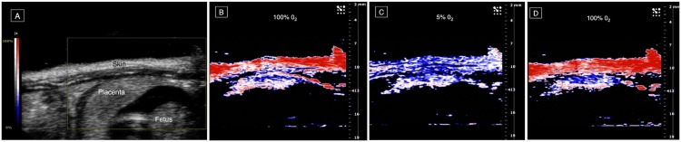
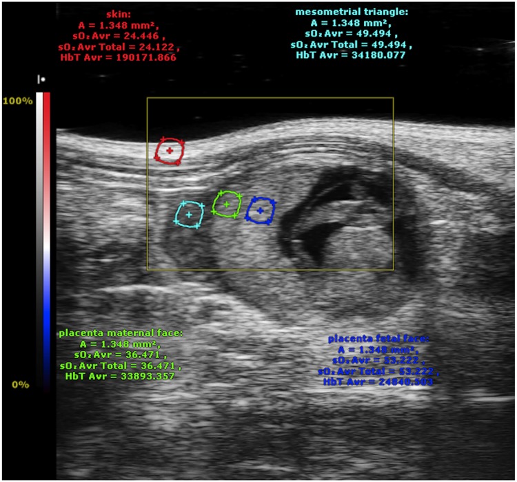
Placentas of fifteen Sprague-Dawley rats were examined on days 14, 17, and 20 of pregnancy with a PA imaging system coupled to high-resolution ultrasound imaging. Pregnant rats were successively exposed to hyperoxygenated and hypoxic conditions by changing the oxygen concentration in inhaled gas. Tissue oxygen saturation was quantitatively analyzed by real-time PA imaging in the skin and 3 regions of the placenta. All procedures were performed in accordance with applicable ethical guidelines and approved by the animal care committee.
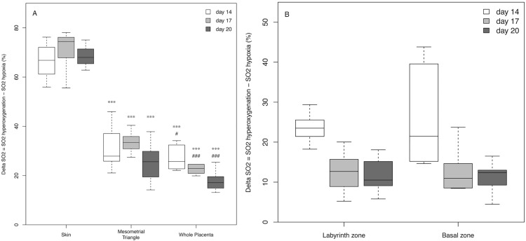
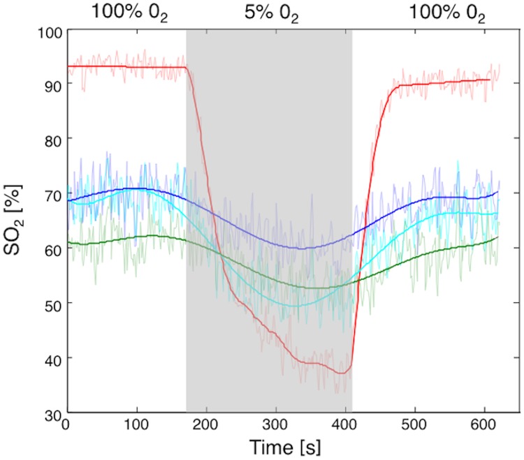
Maternal hypoxia was associated with significantly greater decrease in blood oxygen saturation (ΔO2 Saturation) in the skin (70.74% ±7.65) than in the mesometrial triangle (32.66% ±5.75) or other placental areas (labyrinth: 18.58% ± 6.61; basal zone: 13.13% ±5.72) on different days of pregnancy (P<0.001). ΔO2 Saturation did not differ significantly between the labyrinth, the basal zone, and the decidua. After the period of hypoxia, maternal hyperoxygenation led to a significant rise in oxygen saturation, which returned to its initial values in the different placental regions (P<0.001).
PA imaging enables the variation of blood oxygen saturation to be monitored in the placenta during maternal hypoxia or hyperoxygenation. This first preclinical study suggests that the placenta plays an important role in protecting the fetus against maternal hypoxia.
本临床前研究旨在通过对妊娠不同天数的孕鼠进行实时光声(PA)成像来评估胎盘氧合情况,并明确母体缺氧和高氧状态下胎盘氧饱和度的变化。
使用与高分辨率超声成像耦合的PA成像系统,对15只斯普拉格-道利大鼠在妊娠第14、17和20天的胎盘进行检查。通过改变吸入气体中的氧浓度,使孕鼠依次暴露于高氧和低氧环境。通过实时PA成像对皮肤和胎盘的3个区域的组织氧饱和度进行定量分析。所有操作均按照适用的伦理准则进行,并获得动物护理委员会的批准。
在妊娠不同天数,母体缺氧时皮肤的血氧饱和度下降幅度(ΔO2饱和度)(70.74%±7.65)显著大于子宫系膜三角区(32.66%±5.75)或胎盘其他区域(迷路:18.58%±6.61;基底层:13.13%±5.72)(P<0.001)。迷路、基底层和蜕膜之间的ΔO2饱和度差异不显著。缺氧期过后,母体高氧导致氧饱和度显著上升,不同胎盘区域的氧饱和度恢复到初始值(P<0.001)。
PA成像能够监测母体缺氧或高氧期间胎盘血氧饱和度的变化。这项首次临床前研究表明,胎盘在保护胎儿免受母体缺氧影响方面发挥着重要作用。