Department of Biomedical Engineering, Tulane University, 500 Lindy Boggs Center, New Orleans, LA, 70118, USA.
Department of Biomedical Engineering, Tulane University, 500 Lindy Boggs Center, New Orleans, LA, 70118, USA.
Placenta. 2022 Aug;126:46-53. doi: 10.1016/j.placenta.2022.06.006. Epub 2022 Jun 21.
There is a lack of effective therapeutic interventions for preeclampsia. A central factor in the etiology of the disease is the development of placental hypoxia due to abnormal vascular remodeling. However, methods to assess the impact of potential therapies on placental growth and remodeling are currently lacking. Here, we develop and validate ultrasound-guided photoacoustic imaging methods to monitor the placental response to therapeutic intervention. Establishing non-invasive tools to image placental function opens up previously unachievable understandings of placental therapeutic response.
Studies were performed in the reduced uterine perfusion pressure (RUPP) rat model of preeclampsia. Preclinical research has identified tempol, a superoxide dismutase mimetic, and the phosphodiesterase inhibitor sildenafil as potential therapeutics for preeclampsia, as both improve in vivo maternal outcomes. PA images of the placental environment were acquired in RUPP rats receiving tempol (n = 8) or sildenafil (n = 8) to assess the longitudinal effects of treatment on placental oxygenation and vascular remodeling. Imaging measurements were validated with ex vivo histological analysis.
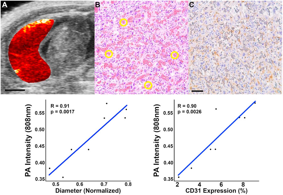
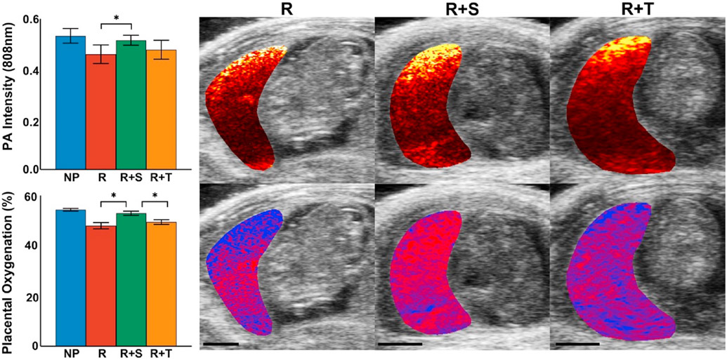
Spectral photoacoustic imaging non-invasively measured placental hypoxia and impaired vascular growth two days after the RUPP procedure was implemented. Sildenafil significantly improved (p < 0.05) placental oxygenation and promoted vascular remodeling in RUPP animals, while RUPP animals treated with tempol had a diminished placental therapeutic response.
We demonstrate that photoacoustic imaging provides in vivo measures of placental oxygenation and vascular remodeling, a previously unobtainable assessment of preeclamptic therapeutic response. These imaging tools have tremendous potential to accelerate the search for effective therapies for preeclampsia.
目前,子痫前期缺乏有效的治疗干预手段。该疾病的一个重要病因是由于血管重塑异常导致胎盘缺氧。然而,目前还缺乏评估潜在治疗方法对胎盘生长和重塑影响的方法。在这里,我们开发并验证了超声引导的光声成像方法来监测胎盘对治疗干预的反应。建立非侵入性的工具来对胎盘功能成像,将为胎盘治疗反应提供以前无法获得的理解。
本研究在子宫灌注压降低(RUPP)的子痫前期大鼠模型中进行。临床前研究已经确定了作为子痫前期潜在治疗药物的 Tempol(一种超氧化物歧化酶模拟物)和磷酸二酯酶抑制剂西地那非,因为它们都能改善体内的母婴结局。在接受 Tempol(n=8)或西地那非(n=8)治疗的 RUPP 大鼠中获取胎盘环境的光声图像,以评估治疗对胎盘氧合和血管重塑的纵向影响。通过离体组织学分析对成像测量结果进行验证。
光谱光声成像非侵入性地测量了 RUPP 手术后两天胎盘缺氧和血管生长受损的情况。西地那非显著改善(p<0.05)了 RUPP 动物的胎盘氧合,并促进了 RUPP 动物的血管重塑,而接受 Tempol 治疗的 RUPP 动物的胎盘治疗反应减弱。
我们证明了光声成像提供了胎盘氧合和血管重塑的体内测量方法,这是对子痫前期治疗反应的以前无法获得的评估。这些成像工具具有巨大的潜力,可以加速寻找有效的子痫前期治疗方法。