Morita Flavio Hiroshi Ananias, Bernardo Wanderley Marques, Ide Edson, Rocha Rodrigo Silva Paula, Aquino Julio Cesar Martins, Minata Mauricio Kazuyoshi, Yamazaki Kendi, Marques Sergio Barbosa, Sakai Paulo, de Moura Eduardo Guimarães Hourneaux
Gastrointestinal Endoscopy at University of Sao Paulo, Rua Capote Valente n 671, Pinheiros, São Paulo, Zipcode 05409-002, Brazil.
University of Sao Paulo, Rua Maria Vidal 124, Perdizes, São Paulo, SP, CEP 01253-040, Brazil.
BMC Cancer. 2017 Jan 13;17(1):54. doi: 10.1186/s12885-016-3011-9.
In the early stage esophageal cancer, changes in the mucosa are subtle and pass unnoticed in endoscopic examinations using white light. To increase sensitivity, chromoscopy with Lugol's solution has been used. Technological advancements have led to the emergence of virtual methods of endoscopic chromoscopy, including narrow band imaging (NBI). NBI enhances the relief of the mucosa and the underlying vascular pattern, providing greater convenience without the risks inherent to the use of vital dye. The purpose of this systematic review and meta-analysis was to evaluate the ability of NBI to diagnose squamous cell carcinoma of the esophagus and to compare it to chromoscopy with Lugol's solution.
This systematic review included all studies comparing the diagnostic accuracy of NBI and Lugol chromoendoscopy performed to identify high-grade dysplasia and/or squamous cell carcinoma in the esophagus. In the meta-analysis, we calculated and demonstrated sensitivity, specificity, and positive and negative likelihood values in forest plots. We also determined summary receiver operating characteristic (sROC) curves and estimates of the areas under the curves for both per-patient and per-lesion analysis.
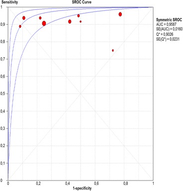
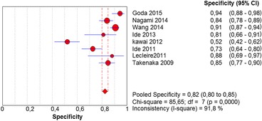
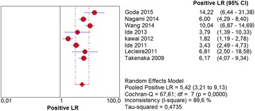
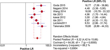
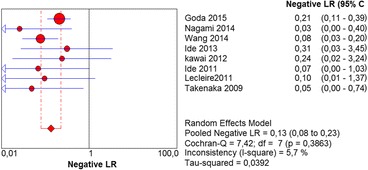
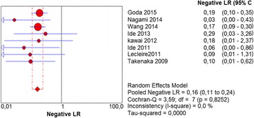
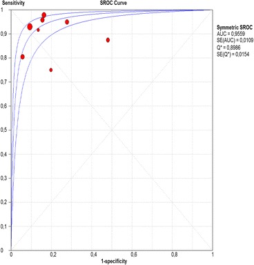
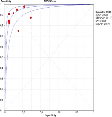
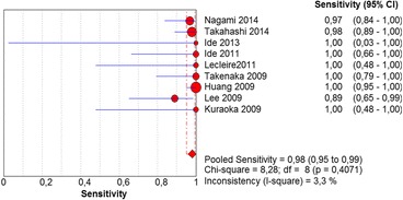
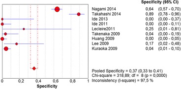
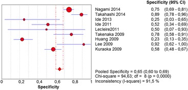
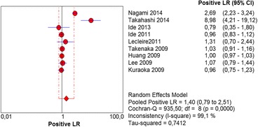
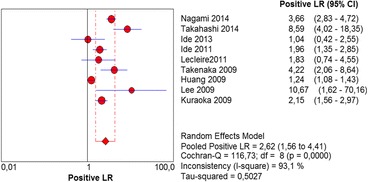
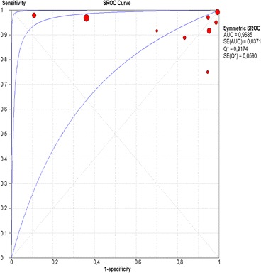
The initial search identified 7079 articles. Of these, 18 studies were included in the systematic review and 12 were used in the meta-analysis, for a total of 1911 patients. In per-patient and per-lesion analysis, the sensitivity, specificity, and positive and negative likelihood values for Lugol chromoendoscopy were 92% and 98, 82 and 37%, 5.42 and 1.4, and 0.13 and 0.39, respectively, and for NBI were 88 and 94%, 88 and 65%, 8.32 and 2.62, and 0.16 and 0.12, respectively. There was a statistically significant difference in only specificity values, in which case NBI was superior to Lugol chromoendoscopy in both analyses. In the per-patient analysis, the area under the sROC curve for Lugol chromoendoscopy was 0.9559. In the case of NBI, this value was 0.9611; in the per-lesion analysis, this number was 0.9685 and 0.9587, respectively.
NBI was adequate in evaluating the esophagus in order to diagnose high-grade dysplasia and squamous cell carcinoma. In the differentiation of those disorders from other esophageal mucosa alterations, the NBI was shown to be superior than Lugol.
在早期食管癌中,黏膜变化细微,在白光内镜检查中易被忽视。为提高敏感性,已采用卢戈氏碘液染色法。技术进步催生了内镜染色的虚拟方法,包括窄带成像(NBI)。NBI可增强黏膜及深层血管形态的对比度,使用方便,且无使用活体染料的固有风险。本系统评价和荟萃分析旨在评估NBI诊断食管鳞状细胞癌的能力,并与卢戈氏碘液染色法进行比较。
本系统评价纳入了所有比较NBI和卢戈氏碘液染色内镜检查诊断食管高级别异型增生和/或鳞状细胞癌准确性的研究。在荟萃分析中,我们计算并在森林图中展示了敏感性、特异性以及阳性和阴性似然值。我们还确定了汇总受试者工作特征(sROC)曲线,并对每位患者和每个病变的分析计算曲线下面积估计值。
初步检索共识别出7079篇文章。其中,18项研究纳入系统评价,12项用于荟萃分析,共计1911例患者。在每位患者和每个病变的分析中,卢戈氏碘液染色内镜检查的敏感性、特异性、阳性和阴性似然值分别为92%和98、82%和37%、5.42和1.4、0.13和0.39,而NBI的相应值分别为88%和94%、88%和65%、8.32和2.62、0.16和0.12。仅特异性值存在统计学显著差异,在两种分析中,NBI的特异性均优于卢戈氏碘液染色内镜检查。在每位患者的分析中,卢戈氏碘液染色内镜检查的sROC曲线下面积为0.9559。NBI的该值为0.9611;在每个病变的分析中,该数值分别为0.9685和0.958