Saha Banishree, Kodys Karen, Szabo Gyongyi
Department of Medicine, University of Massachusetts Medical School, Worcester, Massachusetts.
Cell Mol Gastroenterol Hepatol. 2016 Jan 8;2(3):302-316.e8. doi: 10.1016/j.jcmgh.2015.12.005. eCollection 2016 May.
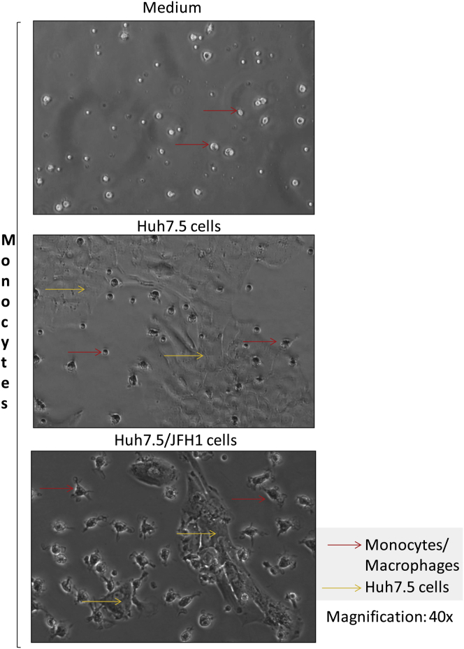
BACKGROUND & AIMS: Monocyte and macrophage (MΦ) activation contributes to the pathogenesis of chronic hepatitis C virus (HCV) infection. Disease pathogenesis is regulated by both liver-resident MΦs and monocytes recruited as precursors of MΦs into the damaged liver. Monocytes differentiate into M1 (classic/proinflammatory) or M2 (alternative/anti-inflammatory) polarized MΦs in response to tissue microenvironment. We hypothesized that HCV-infected hepatoma cells (infected with Japanese fulminant hepatitis-1 [Huh7.5/JFH-1]) induce monocyte differentiation into polarized MΦs.
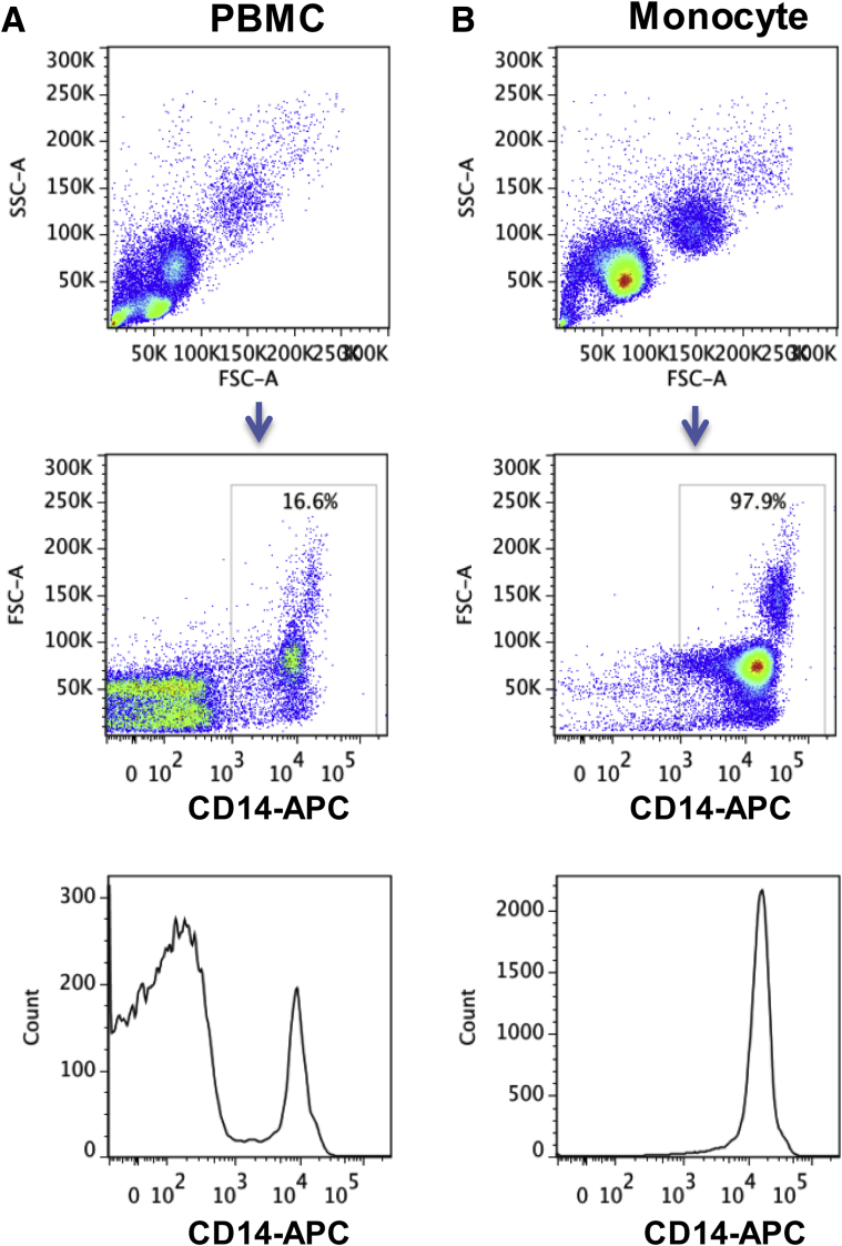
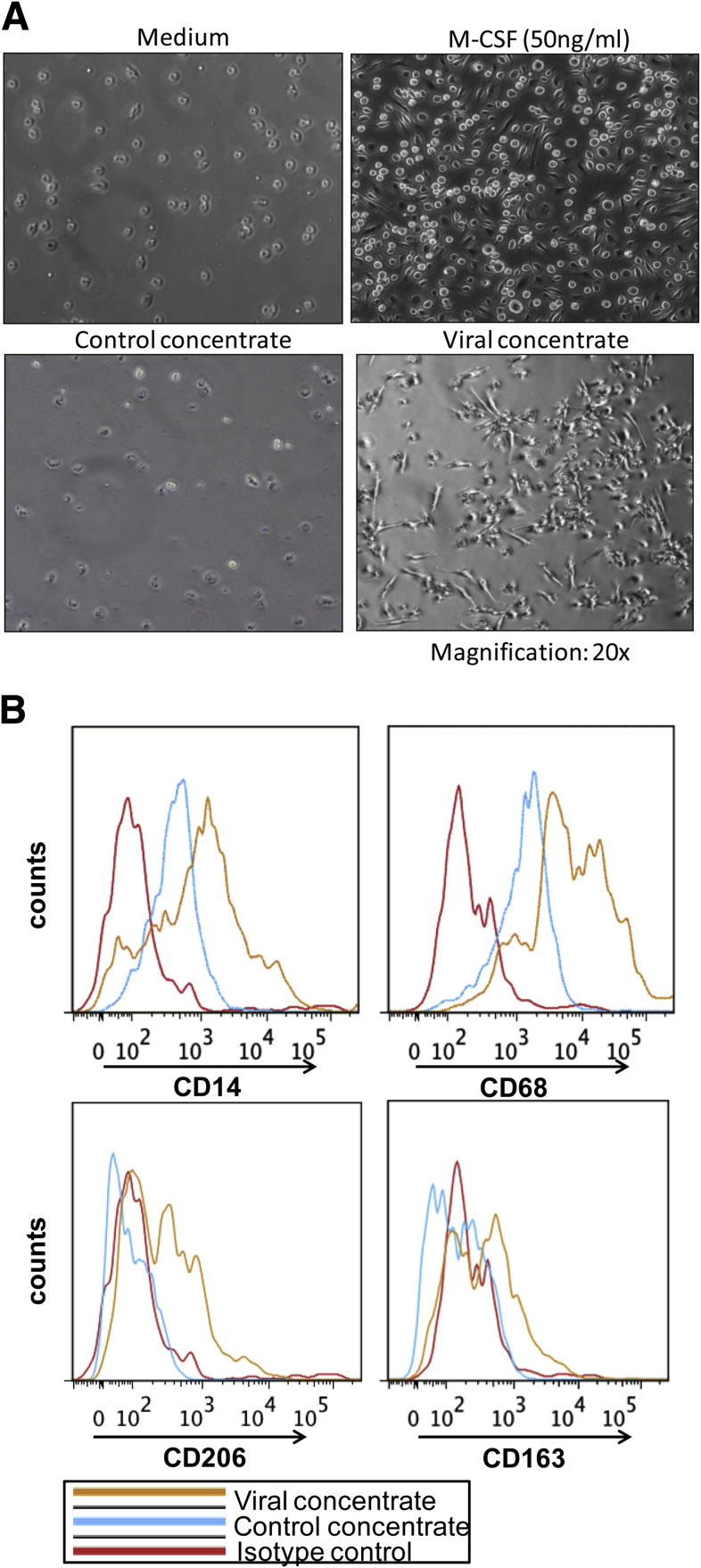
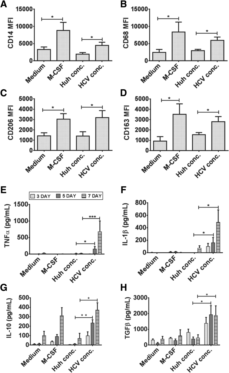
Healthy human monocytes were co-cultured with Huh7.5/JFH-1 cells or cell-free virus for 7 days and analyzed for MΦ markers and cytokine levels. A similar analysis was performed on circulating monocytes and liver MΦs from HCV-infected patients and controls.
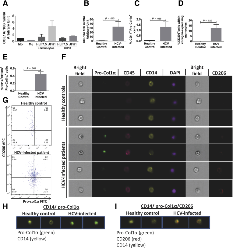
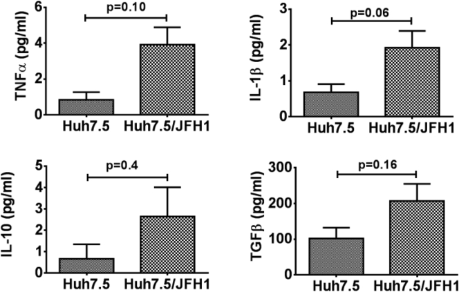
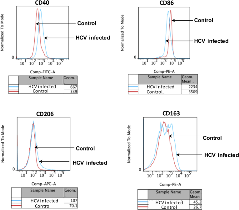
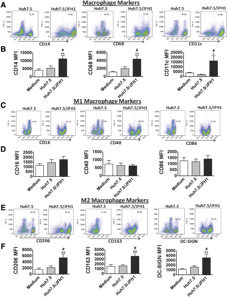
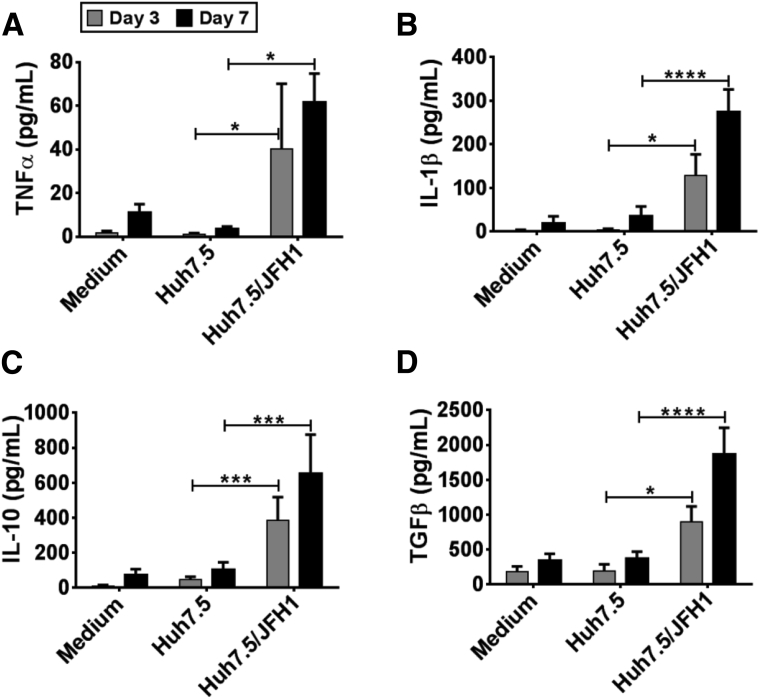
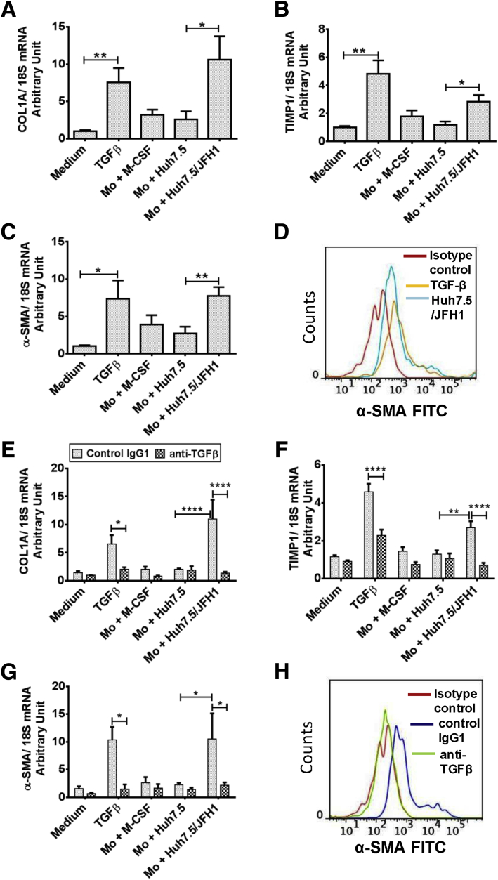
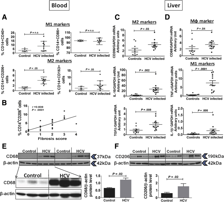
Huh7.5/JFH-1 cells induced monocytes to differentiate into MΦs with increased expression of CD14 and CD68. HCV-MΦs showed M2 surface markers (CD206, CD163, and Dendritic cell-specific intercellular adhesion molecule-3-grabbing non-integrin (DC-SIGN)) and produced both proinflammatory and anti-inflammatory cytokines. HCV-induced early interleukin 1β production promoted transforming growth factor (TGF)β production and MΦ polarization to an M2 phenotype. TGF-β secreted by M2-MΦ led to hepatic stellate cell activation indicated by increased expression of collagen, tissue inhibitor of metalloproteinase 1, and α-smooth muscle actin. In vivo, we observed a significant increase in M2 marker (CD206) expression on circulating monocytes and in the liver of chronic HCV-infected patients. Furthermore, we observed the presence of a unique collagen-expressing CD14CD206 monocyte population in HCV patients that correlated with liver fibrosis.
We show an important role for HCV in induction of monocyte differentiation into MΦs with a mixed M1/M2 cytokine profile and M2 surface phenotype that promote stellate cell activation via TGF-β. We also identified circulating monocytes expressing M2 marker and collagen in chronic HCV infection that can be explored as a biomarker.
单核细胞和巨噬细胞(MΦ)激活参与慢性丙型肝炎病毒(HCV)感染的发病机制。疾病发病机制受肝内驻留MΦs和作为MΦs前体募集到受损肝脏中的单核细胞调控。单核细胞根据组织微环境分化为M1(经典/促炎)或M2(替代/抗炎)极化MΦs。我们推测HCV感染的肝癌细胞(感染日本暴发性肝炎-1 [Huh7.5/JFH-1])诱导单核细胞分化为极化MΦs。
将健康人单核细胞与Huh7.5/JFH-1细胞或无细胞病毒共培养7天,并分析MΦ标志物和细胞因子水平。对HCV感染患者和对照组的循环单核细胞和肝脏MΦs进行类似分析。
Huh7.5/JFH-1细胞诱导单核细胞分化为MΦs,CD14和CD68表达增加。HCV-MΦs表现出M2表面标志物(CD206、CD163和树突状细胞特异性细胞间粘附分子3结合非整合素(DC-SIGN)),并产生促炎和抗炎细胞因子。HCV诱导的早期白细胞介素1β产生促进转化生长因子(TGF)β产生以及MΦ极化至M2表型。M2-MΦ分泌的TGF-β导致肝星状细胞激活,表现为胶原蛋白、金属蛋白酶组织抑制剂1和α-平滑肌肌动蛋白表达增加。在体内,我们观察到慢性HCV感染患者循环单核细胞和肝脏中M2标志物(CD206)表达显著增加。此外,我们在HCV患者中观察到存在独特的表达胶原蛋白的CD14CD206单核细胞群体,其与肝纤维化相关。
我们表明HCV在诱导单核细胞分化为具有混合M1/M2细胞因子谱和M2表面表型的MΦs中起重要作用,这些MΦs通过TGF-β促进星状细胞激活。我们还在慢性HCV感染中鉴定出表达M2标志物和胶原蛋白的循环单核细胞,可将其作为生物标志物进行研究。