• 文献检索
  • 文档翻译
  • 深度研究
  • 学术资讯
  • Suppr Zotero 插件Zotero 插件
  • 邀请有礼
  • 套餐&价格
  • 历史记录
应用&插件
Suppr Zotero 插件Zotero 插件浏览器插件Mac 客户端Windows 客户端微信小程序
定价
高级版会员购买积分包购买API积分包
服务
文献检索文档翻译深度研究API 文档MCP 服务
关于我们
关于 Suppr公司介绍联系我们用户协议隐私条款
关注我们

Suppr 超能文献

核心技术专利:CN118964589B侵权必究
粤ICP备2023148730 号-1Suppr @ 2026

文献检索

告别复杂PubMed语法,用中文像聊天一样搜索,搜遍4000万医学文献。AI智能推荐,让科研检索更轻松。

立即免费搜索

文件翻译

保留排版,准确专业,支持PDF/Word/PPT等文件格式,支持 12+语言互译。

免费翻译文档

深度研究

AI帮你快速写综述,25分钟生成高质量综述,智能提取关键信息,辅助科研写作。

立即免费体验

ultraLM和miniLM:用于在相关光电子显微镜中智能追踪荧光细胞的定位工具。

ultraLM and miniLM: Locator tools for smart tracking of fluorescent cells in correlative light and electron microscopy.

作者信息

Brama Elisabeth, Peddie Christopher J, Wilkes Gary, Gu Yan, Collinson Lucy M, Jones Martin L

机构信息

Electron Microscopy Science Technology Platform, The Francis Crick Institute, London, UK.

出版信息

Wellcome Open Res. 2016 Dec 13;1:26. doi: 10.12688/wellcomeopenres.10299.1.

DOI:10.12688/wellcomeopenres.10299.1
PMID:28090593
原文链接:https://pmc.ncbi.nlm.nih.gov/articles/PMC5234702/
Abstract

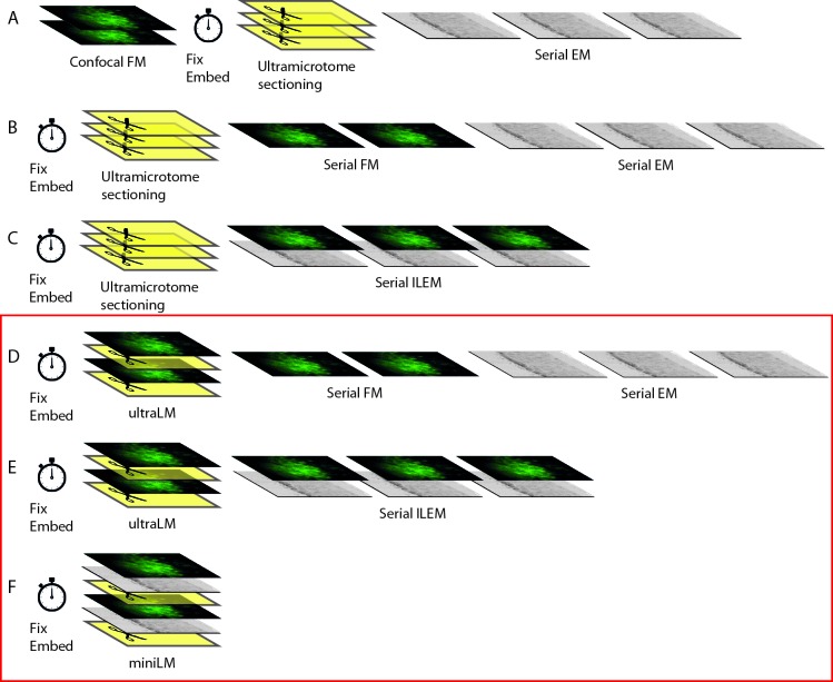
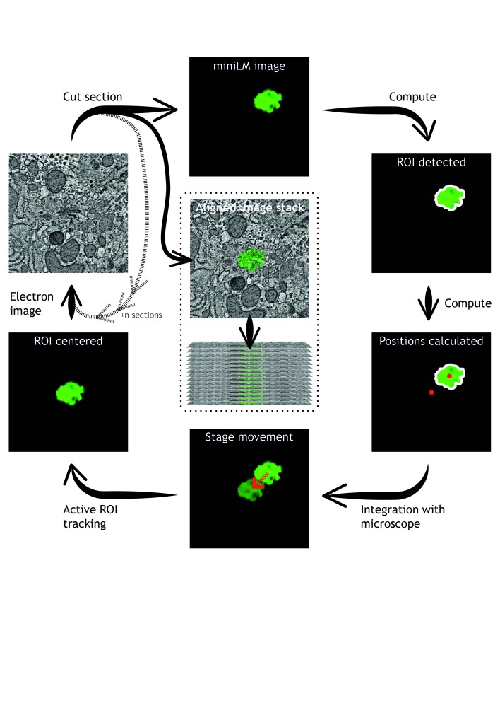
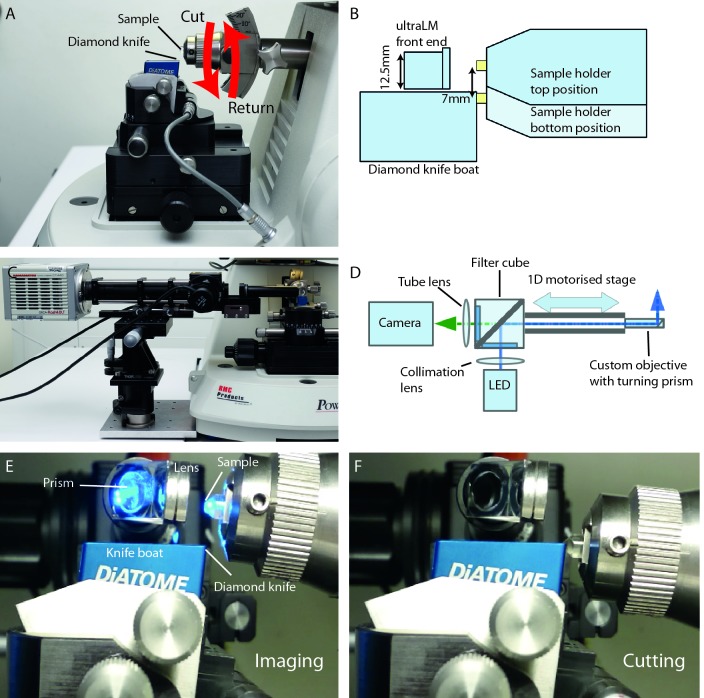
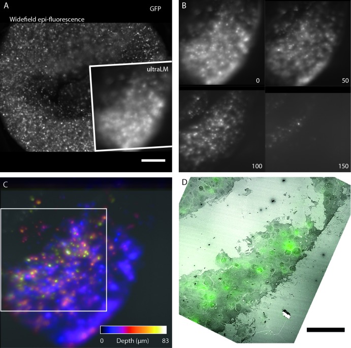
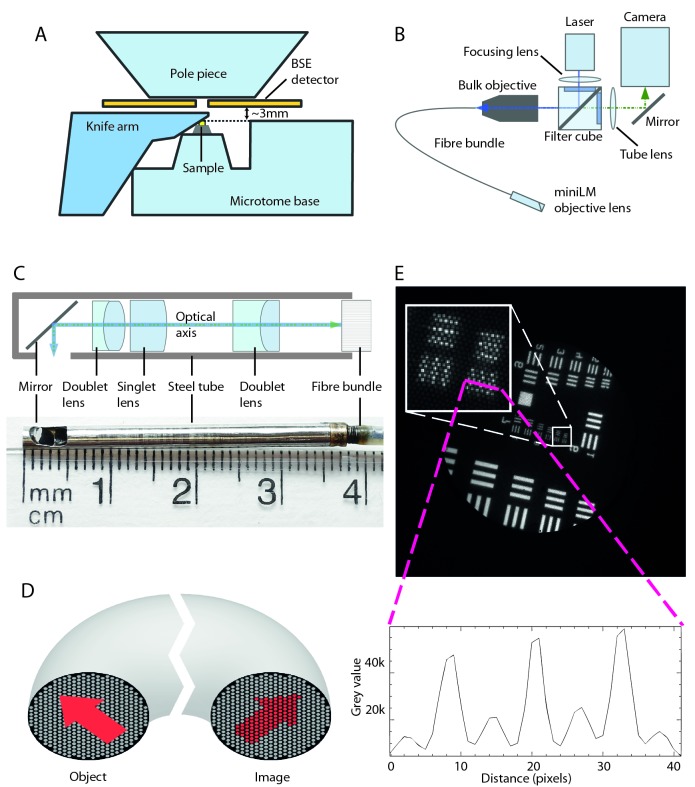
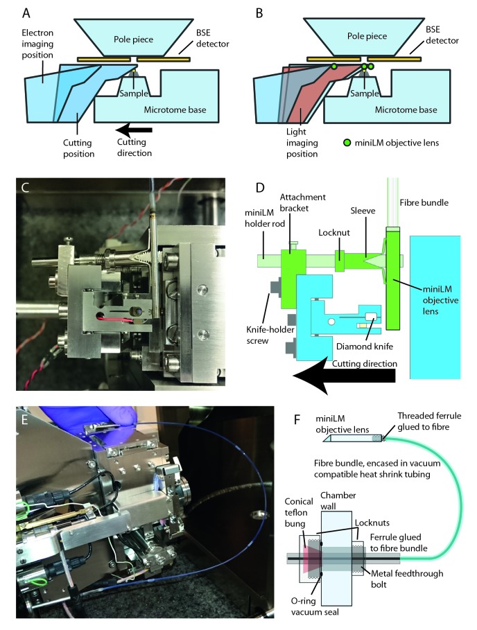
In-resin fluorescence (IRF) protocols preserve fluorescent proteins in resin-embedded cells and tissues for correlative light and electron microscopy, aiding interpretation of macromolecular function within the complex cellular landscape. Dual-contrast IRF samples can be imaged in separate fluorescence and electron microscopes, or in dual-modality integrated microscopes for high resolution correlation of fluorophore to organelle. IRF samples also offer a unique opportunity to automate correlative imaging workflows. Here we present two new locator tools for finding and following fluorescent cells in IRF blocks, enabling future automation of correlative imaging. The ultraLM is a fluorescence microscope that integrates with an ultramicrotome, which enables 'smart collection' of ultrathin sections containing fluorescent cells or tissues for subsequent transmission electron microscopy or array tomography. The miniLM is a fluorescence microscope that integrates with serial block face scanning electron microscopes, which enables 'smart tracking' of fluorescent structures during automated serial electron image acquisition from large cell and tissue volumes.

摘要

树脂内荧光(IRF)方案可在树脂包埋的细胞和组织中保存荧光蛋白,用于相关光镜和电镜检查,有助于在复杂的细胞环境中解释大分子功能。双对比IRF样本可在单独的荧光显微镜和电子显微镜中成像,也可在双模态集成显微镜中成像,以实现荧光团与细胞器的高分辨率关联。IRF样本还为自动化相关成像工作流程提供了独特的机会。在这里,我们展示了两种新的定位工具,用于在IRF块中查找和跟踪荧光细胞,从而实现未来相关成像的自动化。ultraLM是一种与超薄切片机集成的荧光显微镜,可对含有荧光细胞或组织的超薄切片进行“智能采集”,以便后续进行透射电子显微镜检查或阵列断层扫描。miniLM是一种与连续块面扫描电子显微镜集成的荧光显微镜,可在从大细胞和组织体积自动采集连续电子图像期间对荧光结构进行“智能跟踪”。

https://cdn.ncbi.nlm.nih.gov/pmc/blobs/cddd/5446011/6ca30ca7e901/wellcomeopenres-1-11092-g0007.jpg
https://cdn.ncbi.nlm.nih.gov/pmc/blobs/cddd/5446011/17b7b60e9255/wellcomeopenres-1-11092-g0000.jpg
https://cdn.ncbi.nlm.nih.gov/pmc/blobs/cddd/5446011/f554afcf912a/wellcomeopenres-1-11092-g0001.jpg
https://cdn.ncbi.nlm.nih.gov/pmc/blobs/cddd/5446011/1cc4c92f9d53/wellcomeopenres-1-11092-g0002.jpg
https://cdn.ncbi.nlm.nih.gov/pmc/blobs/cddd/5446011/706b89a31e21/wellcomeopenres-1-11092-g0003.jpg
https://cdn.ncbi.nlm.nih.gov/pmc/blobs/cddd/5446011/db3d00e415be/wellcomeopenres-1-11092-g0004.jpg
https://cdn.ncbi.nlm.nih.gov/pmc/blobs/cddd/5446011/4e3918fb1834/wellcomeopenres-1-11092-g0005.jpg
https://cdn.ncbi.nlm.nih.gov/pmc/blobs/cddd/5446011/84b29af3cfb8/wellcomeopenres-1-11092-g0006.jpg
https://cdn.ncbi.nlm.nih.gov/pmc/blobs/cddd/5446011/6ca30ca7e901/wellcomeopenres-1-11092-g0007.jpg

相似文献

1
ultraLM and miniLM: Locator tools for smart tracking of fluorescent cells in correlative light and electron microscopy.ultraLM和miniLM:用于在相关光电子显微镜中智能追踪荧光细胞的定位工具。
Wellcome Open Res. 2016 Dec 13;1:26. doi: 10.12688/wellcomeopenres.10299.1.
2
A workflow for 3D-CLEM investigating liver tissue.用于研究肝组织的 3D-CLEM 工作流程。
J Microsc. 2021 Mar;281(3):231-242. doi: 10.1111/jmi.12967. Epub 2020 Oct 27.
3
Automated detection of fluorescent cells in in-resin fluorescence sections for integrated light and electron microscopy.在树脂荧光切片中自动检测荧光细胞,用于整合光镜和电镜。
J Microsc. 2018 Jul;271(1):109-119. doi: 10.1111/jmi.12700. Epub 2018 Apr 26.
4
Correlative super-resolution fluorescence and electron microscopy using conventional fluorescent proteins in vacuo.在真空中使用传统荧光蛋白的相关超分辨率荧光和电子显微镜技术。
J Struct Biol. 2017 Aug;199(2):120-131. doi: 10.1016/j.jsb.2017.05.013. Epub 2017 May 30.
5
Multimodal Hierarchical Imaging of Serial Sections for Finding Specific Cellular Targets within Large Volumes.用于在大体积样本中寻找特定细胞靶点的连续切片多模态分层成像
J Vis Exp. 2018 Mar 20(133):57059. doi: 10.3791/57059.
6
Integrated light and scanning electron microscopy of GFP-expressing cells.绿色荧光蛋白表达细胞的光学与扫描电子显微镜联合成像
Methods Cell Biol. 2014;124:363-89. doi: 10.1016/B978-0-12-801075-4.00017-3.
7
Correlative and integrated light and electron microscopy of in-resin GFP fluorescence, used to localise diacylglycerol in mammalian cells.用于在哺乳动物细胞中定位二酰基甘油的树脂包埋绿色荧光蛋白(GFP)荧光的相关及整合光镜和电镜技术。
Ultramicroscopy. 2014 Aug;143(100):3-14. doi: 10.1016/j.ultramic.2014.02.001. Epub 2014 Feb 22.
8
Advanced cryo-tomography workflow developments - correlative microscopy, milling automation and cryo-lift-out.先进的冷冻电子断层扫描工作流程发展——相关显微镜技术、铣削自动化和冷冻提取技术。
J Microsc. 2021 Feb;281(2):112-124. doi: 10.1111/jmi.12939. Epub 2020 Jul 2.
9
Preserving the photoswitching ability of standard fluorescent proteins for correlative in-resin super-resolution and electron microscopy.保留标准荧光蛋白的光开关能力用于树脂内相关超分辨率和电子显微镜观察。
Methods Cell Biol. 2017;140:49-67. doi: 10.1016/bs.mcb.2017.04.001. Epub 2017 May 5.
10
Reactive oxygen FIB spin milling enables correlative workflow for 3D super-resolution light microscopy and serial FIB/SEM of cultured cells.活性氧纤维铣削实现了用于培养细胞的 3D 超分辨率光显微镜和连续纤维切割/扫描电镜的相关工作流程。
Sci Rep. 2021 Jun 23;11(1):13162. doi: 10.1038/s41598-021-92608-y.

引用本文的文献

1
CELS-3D-Cutting edge light source for exciting fluorescence in microtome-based 3D microscopy and targeted correlative microscopy.CELS-3D——用于基于切片机的三维显微镜和靶向相关显微镜中激发荧光的前沿光源。
J Anat. 2025 May;246(5):782-797. doi: 10.1111/joa.14164. Epub 2024 Nov 27.
2
Crosshair, semi-automated targeting for electron microscopy with a motorised ultramicrotome.Crosshair,用于带自动马达的超薄切片机的电子显微镜半自动靶向。
Elife. 2022 Nov 15;11:e80899. doi: 10.7554/eLife.80899.
3
One for All, All for One: A Close Look at In-Resin Fluorescence Protocols for CLEM.

本文引用的文献

1
Comparing homologous microscopic sections from multiple embryos using HREM.使用高分辨率电子显微镜(HREM)比较多个胚胎的同源显微切片。
Dev Biol. 2016 Jul 1;415(1):1-5. doi: 10.1016/j.ydbio.2016.05.011. Epub 2016 May 18.
2
Biophotonic endoscopy: a review of clinical research techniques for optical imaging and sensing of early gastrointestinal cancer.生物光子内镜检查:早期胃肠道癌光学成像与传感临床研究技术综述
Endosc Int Open. 2015 Oct;3(5):E380-92. doi: 10.1055/s-0034-1392513. Epub 2015 Sep 8.
3
Correlative in-resin super-resolution and electron microscopy using standard fluorescent proteins.
我为人人,人人为我:深入探究用于CLEM的树脂内荧光方案。
Front Cell Dev Biol. 2022 Jun 30;10:866472. doi: 10.3389/fcell.2022.866472. eCollection 2022.
4
Correlative Organelle Microscopy: Fluorescence Guided Volume Electron Microscopy of Intracellular Processes.相关细胞器显微镜检查:细胞内过程的荧光引导体积电子显微镜检查
Front Cell Dev Biol. 2022 Apr 11;10:829545. doi: 10.3389/fcell.2022.829545. eCollection 2022.
5
A workflow for streamlined acquisition and correlation of serial regions of interest in array tomography.一种用于简化采集和关联面探术的连续感兴趣区域的工作流程。
BMC Biol. 2021 Jul 30;19(1):152. doi: 10.1186/s12915-021-01072-7.
6
Development of protocols for the first serial block-face scanning electron microscopy (SBF SEM) studies of bone tissue.制定用于骨组织的第一代连续块面扫描电子显微镜 (SBF SEM) 研究的方案。
Bone. 2020 Feb;131:115107. doi: 10.1016/j.bone.2019.115107. Epub 2019 Oct 24.
7
Dedicated Setup for the Photoconversion of Fluorescent Dyes for Functional Electron Microscopy.用于功能电子显微镜荧光染料光转换的专用装置。
Front Cell Neurosci. 2019 Jul 24;13:312. doi: 10.3389/fncel.2019.00312. eCollection 2019.
8
From Light Microscopy to Analytical Scanning Electron Microscopy (SEM) and Focused Ion Beam (FIB)/SEM in Biology: Fixed Coordinates, Flat Embedding, Absolute References.从光学显微镜到生物学中的分析扫描电子显微镜(SEM)和聚焦离子束(FIB)/SEM:固定坐标、平面包埋、绝对参考
Microsc Microanal. 2018 Oct;24(5):526-544. doi: 10.1017/S1431927618015015. Epub 2018 Sep 24.
9
Microtome-integrated microscope system for high sensitivity tracking of in-resin fluorescence in blocks and ultrathin sections for correlative microscopy.用于在包埋块和超薄切片中对树脂内荧光进行高灵敏度追踪以进行相关显微镜检查的切片机集成显微镜系统。
Sci Rep. 2017 Oct 19;7(1):13583. doi: 10.1038/s41598-017-13348-6.
使用标准荧光蛋白的相关树脂内超分辨率和电子显微镜技术。
Sci Rep. 2015 Mar 31;5:9583. doi: 10.1038/srep09583.
4
Advanced methods of microscope control using μManager software.使用μManager软件的高级显微镜控制方法。
J Biol Methods. 2014;1(2). doi: 10.14440/jbm.2014.36.
5
Preface. Correlative light and electron microscopy II.前言。相关光镜与电镜检查II。
Methods Cell Biol. 2014;124:xvii-xviii. doi: 10.1016/B978-0-12-801075-4.09983-3.
6
Integrated light and scanning electron microscopy of GFP-expressing cells.绿色荧光蛋白表达细胞的光学与扫描电子显微镜联合成像
Methods Cell Biol. 2014;124:363-89. doi: 10.1016/B978-0-12-801075-4.00017-3.
7
Imaging ATUM ultrathin section libraries with WaferMapper: a multi-scale approach to EM reconstruction of neural circuits.使用 WaferMapper 对 ATUM 超薄切片文库进行成像:一种用于神经回路 EM 重建的多尺度方法。
Front Neural Circuits. 2014 Jun 27;8:68. doi: 10.3389/fncir.2014.00068. eCollection 2014.
8
Correlative and integrated light and electron microscopy of in-resin GFP fluorescence, used to localise diacylglycerol in mammalian cells.用于在哺乳动物细胞中定位二酰基甘油的树脂包埋绿色荧光蛋白(GFP)荧光的相关及整合光镜和电镜技术。
Ultramicroscopy. 2014 Aug;143(100):3-14. doi: 10.1016/j.ultramic.2014.02.001. Epub 2014 Feb 22.
9
Correlative imaging of fluorescent proteins in resin-embedded plant material.荧光蛋白在树脂包埋植物材料中的相关成像。
Plant Physiol. 2013 Apr;161(4):1595-603. doi: 10.1104/pp.112.212365. Epub 2013 Mar 1.
10
Simultaneous correlative scanning electron and high-NA fluorescence microscopy.同时关联扫描电子显微镜和高数值孔径荧光显微镜。
PLoS One. 2013;8(2):e55707. doi: 10.1371/journal.pone.0055707. Epub 2013 Feb 8.