Institute of Reconstructive Neurobiology, University of Bonn, Sigmund-Freud-Strasse 25, 53127 Bonn, Germany.
Life&Brain GmbH, Sigmund-Freud-Strasse 25, 53127 Bonn, Germany.
Nat Commun. 2017 Jan 19;8:14162. doi: 10.1038/ncomms14162.
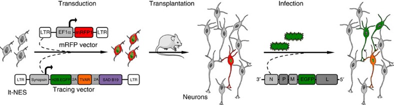
While transplantation represents a key tool for assessing in vivo functionality of neural stem cells and their suitability for neural repair, little is known about the integration of grafted neurons into the host brain circuitry. Rabies virus-based retrograde tracing has developed into a powerful approach for visualizing synaptically connected neurons. Here, we combine this technique with light sheet fluorescence microscopy (LSFM) to visualize transplanted cells and connected host neurons in whole-mouse brain preparations. Combined with co-registration of high-precision three-dimensional magnetic resonance imaging (3D MRI) reference data sets, this approach enables precise anatomical allocation of the host input neurons. Our data show that the same neural donor cell population grafted into different brain regions receives highly orthotopic input. These findings indicate that transplant connectivity is largely dictated by the circuitry of the target region and depict rabies-based transsynaptic tracing and LSFM as efficient tools for comprehensive assessment of host-donor cell innervation.
虽然移植是评估神经干细胞体内功能及其用于神经修复的适宜性的重要工具,但对于移植神经元与宿主大脑回路的整合知之甚少。基于狂犬病病毒的逆行追踪已发展成为可视化突触连接神经元的强大方法。在这里,我们将该技术与光片荧光显微镜(LSFM)相结合,用于可视化整个小鼠脑标本中的移植细胞和连接的宿主神经元。结合高精度三维磁共振成像(3D MRI)参考数据集的配准,该方法能够精确分配宿主输入神经元。我们的数据表明,移植到不同脑区的同一神经供体细胞群接收高度同型的输入。这些发现表明,移植连接在很大程度上取决于靶区的回路,并将狂犬病病毒的转导追踪和 LSFM 描绘为全面评估宿主-供体细胞支配的有效工具。