Singh Dharam, Garg R S, Garg Yashika, Arora Vijinder
Department of Orthopaedics, Govt. Medical College, Amritsar, India.
Department of Pharmacology, E.S.I Dental College, New Delhi, India.
J Orthop Case Rep. 2016 Jul-Aug;6(3):38-39. doi: 10.13107/jocr.2250-0685.494.
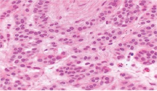
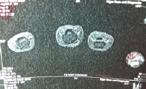
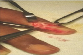
Glomus tumor is a rare benign tumor arising from neuroarterial plexus concentrated beneath the nailbed usually in women of age group 20-40 yrs. The plexus is an arteriovenous anastomosis functioning without intermediary capillary bed, we report a case of glomus tumor affecting the nailbed of left little finger with characteristic periodic, spontaneous excruciating pain and temperature related algesia for the last 6 months. MRI helped in clinching the diagnosis at an early stage. Patient was operated for excision of the tumor making her completely pain free after resection oftumor.
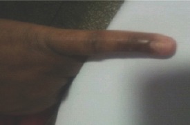
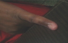
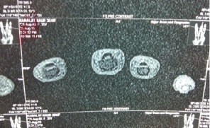
A 40-year-old female patient came in Ortho. OPD with complaints of pain, cold algesia, and point tenderness on the radial side of base of the nailbed of left little finger for the last 6 months. Diagnosis was delayed despite the patient having sought the advice from different clinicians a number of times before coming to us. Pain used to occur on accidental touching of tender spot, cold water immersion; excruciating at times, making the patient faint. On physical examination clinical diagnosis of glomus tumor was made, which was confirmed on MRI as the plain X-ray was non-contributory.
Most of the glomus tumors are benign, which are amenable to cure with complete surgical excision. Rarely, if the lesion exceeds 2 cm malignant transformation of the tumor must be suspected unless proven otherwise. Delay in clinical diagnosis due to dithering on the part of the clinician unnecessarily prolong the suffering in the patient which can be greatly helped by M.R.I in clinching the diagnosis early.
血管球瘤是一种罕见的良性肿瘤,起源于通常在20 - 40岁女性指甲床下集中的神经动脉丛。该丛是一种无中间毛细血管床的动静脉吻合,我们报告一例影响左手小指指甲床的血管球瘤病例,在过去6个月中具有典型的周期性、自发性剧痛和温度相关性疼痛。磁共振成像(MRI)有助于早期确诊。患者接受了肿瘤切除术,术后疼痛完全消失。
一名40岁女性患者因左手小指指甲床基部桡侧疼痛、冷痛觉过敏和压痛点就诊于骨科门诊。在来我们这里之前,患者多次向不同临床医生咨询,但诊断仍被延误。疼痛常在意外触碰压痛点、冷水浸泡时发作;有时剧痛难忍,可使患者昏厥。体格检查做出了血管球瘤的临床诊断,磁共振成像(MRI)证实了这一诊断,因为X线平片无诊断价值。
大多数血管球瘤是良性的,可通过完全手术切除治愈。极少数情况下,如果病变超过2厘米,除非另有证明,否则必须怀疑肿瘤发生恶变。临床医生的犹豫不决导致临床诊断延误,不必要地延长了患者的痛苦,而磁共振成像(MRI)有助于早期确诊,可极大地帮助患者。