Passamonti Luca, Vázquez Rodríguez Patricia, Hong Young T, Allinson Kieren S J, Williamson David, Borchert Robin J, Sami Saber, Cope Thomas E, Bevan-Jones W Richard, Jones P Simon, Arnold Robert, Surendranathan Ajenthan, Mak Elijah, Su Li, Fryer Tim D, Aigbirhio Franklin I, O'Brien John T, Rowe James B
Department of Clinical Neurosciences, University of Cambridge, Cambridge, UK.
Istituto di Bioimmagini e Fisiologia Molecolare, Consiglio Nazionale delle Ricerche, Milano, Italy.
Brain. 2017 Mar 1;140(3):781-791. doi: 10.1093/brain/aww340.
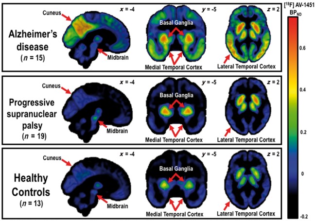
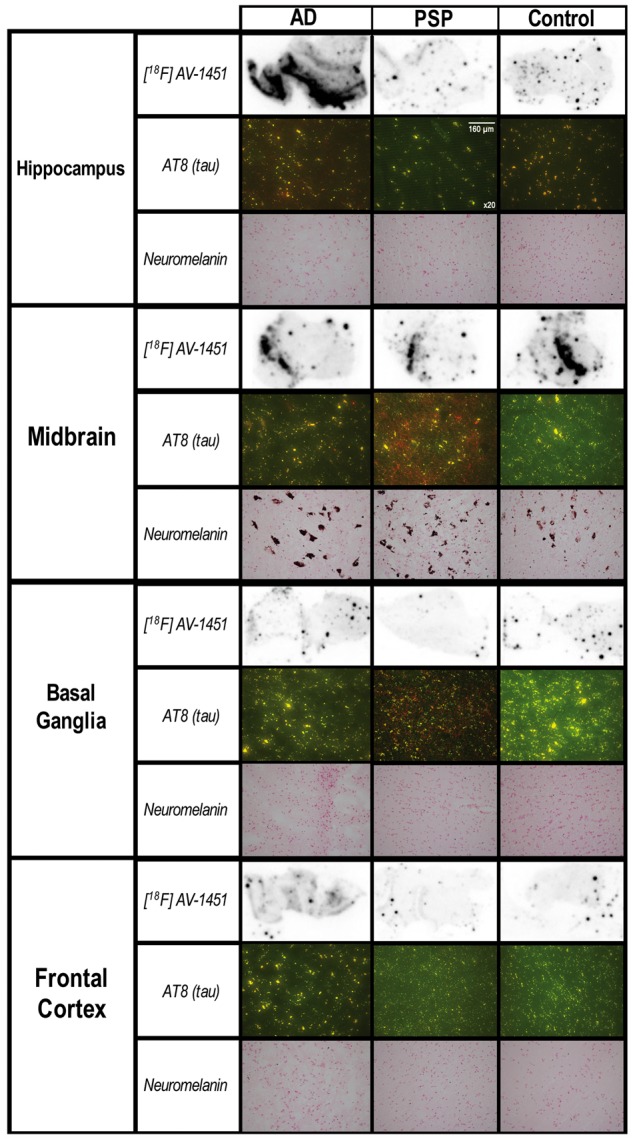
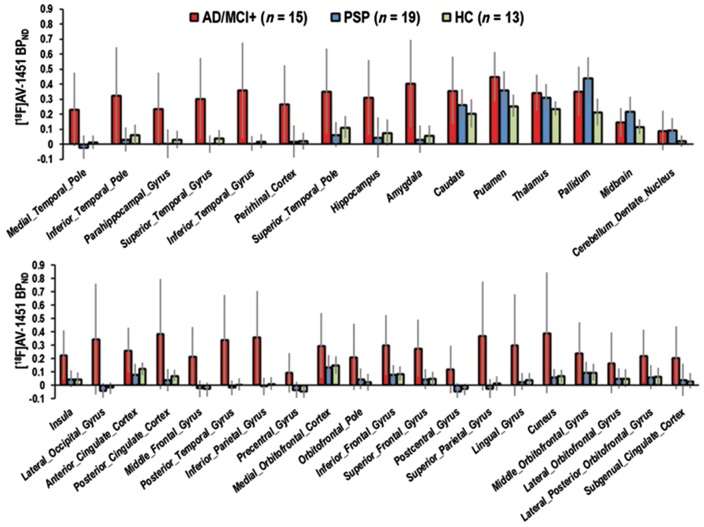
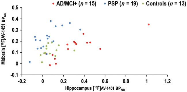
The ability to assess the distribution and extent of tau pathology in Alzheimer's disease and progressive supranuclear palsy in vivo would help to develop biomarkers for these tauopathies and clinical trials of disease-modifying therapies. New radioligands for positron emission tomography have generated considerable interest, and controversy, in their potential as tau biomarkers. We assessed the radiotracer 18F-AV-1451 with positron emission tomography imaging to compare the distribution and intensity of tau pathology in 15 patients with Alzheimer's pathology (including amyloid-positive mild cognitive impairment), 19 patients with progressive supranuclear palsy, and 13 age- and sex-matched controls. Regional analysis of variance and a support vector machine were used to compare and discriminate the clinical groups, respectively. We also examined the 18F-AV-1451 autoradiographic binding in post-mortem tissue from patients with Alzheimer's disease, progressive supranuclear palsy, and a control case to assess the 18F-AV-1451 binding specificity to Alzheimer's and non-Alzheimer's tau pathology. There was increased 18F-AV-1451 binding in multiple regions in living patients with Alzheimer's disease and progressive supranuclear palsy relative to controls [main effect of group, F(2,41) = 17.5, P < 0.0001; region of interest × group interaction, F(2,68) = 7.5, P < 0.00001]. More specifically, 18F-AV-1451 binding was significantly increased in patients with Alzheimer's disease, relative to patients with progressive supranuclear palsy and with control subjects, in the hippocampus and in occipital, parietal, temporal, and frontal cortices (t's > 2.2, P's < 0.04). Conversely, in patients with progressive supranuclear palsy, relative to patients with Alzheimer's disease, 18F-AV-1451 binding was elevated in the midbrain (t = 2.1, P < 0.04); while patients with progressive supranuclear palsy showed, relative to controls, increased 18F-AV-1451 uptake in the putamen, pallidum, thalamus, midbrain, and in the dentate nucleus of the cerebellum (t's > 2.7, P's < 0.02). The support vector machine assigned patients' diagnoses with 94% accuracy. The post-mortem autoradiographic data showed that 18F-AV-1451 strongly bound to Alzheimer-related tau pathology, but less specifically in progressive supranuclear palsy. 18F-AV-1451 binding to the basal ganglia was strong in all groups in vivo. Postmortem histochemical staining showed absence of neuromelanin-containing cells in the basal ganglia, indicating that off-target binding to neuromelanin is an insufficient explanation of 18F-AV-1451 positron emission tomography data in vivo, at least in the basal ganglia. Overall, we confirm the potential of 18F-AV-1451 as a heuristic biomarker, but caution is indicated in the neuropathological interpretation of its binding. Off-target binding may contribute to disease profiles of 18F-AV-1451 positron emission tomography, especially in primary tauopathies such as progressive supranuclear palsy. We suggest that 18F-AV-1451 positron emission tomography is a useful biomarker to assess tau pathology in Alzheimer's disease and to distinguish it from other tauopathies with distinct clinical and pathological characteristics such as progressive supranuclear palsy.
在体内评估阿尔茨海默病和进行性核上性麻痹中tau蛋白病理的分布和程度,将有助于开发针对这些tau蛋白病的生物标志物以及疾病修饰疗法的临床试验。用于正电子发射断层扫描的新型放射性配体,作为tau蛋白生物标志物的潜力引发了广泛关注和争议。我们使用正电子发射断层扫描成像评估放射性示踪剂18F-AV-1451,以比较15例患有阿尔茨海默病病理(包括淀粉样蛋白阳性轻度认知障碍)的患者、19例进行性核上性麻痹患者以及13例年龄和性别匹配的对照者中tau蛋白病理的分布和强度。分别使用方差分析和支持向量机对临床组进行比较和鉴别。我们还检查了来自阿尔茨海默病、进行性核上性麻痹患者及1例对照病例的尸检组织中的18F-AV-1451放射自显影结合情况,以评估18F-AV-1451对阿尔茨海默病和非阿尔茨海默病tau蛋白病理的结合特异性。与对照组相比,患有阿尔茨海默病和进行性核上性麻痹的活体患者多个区域的18F-AV-1451结合增加[组间主效应,F(2,41) = 17.5,P < 0.0001;感兴趣区域×组间交互作用,F(2,68) = 7.5,P < 0.00001]。更具体地说,与进行性核上性麻痹患者和对照受试者相比,阿尔茨海默病患者海马体以及枕叶、顶叶、颞叶和额叶皮质中的18F-AV-1451结合显著增加(t值> 2.2,P值< 0.04)。相反,与阿尔茨海默病患者相比,进行性核上性麻痹患者中脑的18F-AV-1451结合升高(t = 2.1,P < 0.04);而与对照组相比,进行性核上性麻痹患者壳核、苍白球、丘脑、中脑和小脑齿状核中的18F-AV-1451摄取增加(t值> 2.7,P值< 0.02)。支持向量机对患者诊断的准确率为94%。尸检放射自显影数据表明,18F-AV-1451与阿尔茨海默病相关的tau蛋白病理强烈结合,但在进行性核上性麻痹中的特异性较低。在体内所有组中,18F-AV-1451与基底神经节的结合都很强。尸检组织化学染色显示基底神经节中不含神经黑色素的细胞,这表明至少在基底神经节中,与神经黑色素的非靶向结合不足以解释体内18F-AV-1451正电子发射断层扫描数据。总体而言,我们证实了18F-AV-1451作为启发式生物标志物的潜力,但在对其结合进行神经病理学解释时需谨慎。非靶向结合可能导致18F-AV-1451正电子发射断层扫描的疾病特征,尤其是在原发性tau蛋白病如进行性核上性麻痹中。我们认为,18F-AV-1451正电子发射断层扫描是评估阿尔茨海默病中tau蛋白病理并将其与具有不同临床和病理特征的其他tau蛋白病(如进行性核上性麻痹)区分开来的有用生物标志物。