Nasto Luigi Aurelio, Muquit Samiul, Perez-Romera Ana Belen, Mehdian Hossein
The Centre for Spinal Studies and Surgery, Queen's Medical Centre, Nottingham University Hospitals NHS Trust, Derby Road, Nottingham, NG7 2UH, UK.
J Orthop Traumatol. 2017 Jun;18(2):135-143. doi: 10.1007/s10195-016-0440-9. Epub 2017 Jan 25.
Standard laminectomy for treatment of cervical myelopathy is associated with secondary instability and kyphosis, while laminectomy combined with fusion puts adjacent segments at risk of degeneration. Single- and double-door laminoplasty techniques have been developed to overcome these limitations. More recently, complications related to bone graft dislodgment have fostered development of hardware-augmented laminoplasty techniques. The aim of this study is to review the clinical safety and effectiveness of a newly developed technique of instrumented French-door laminoplasty for treatment of cervical myelopathy.
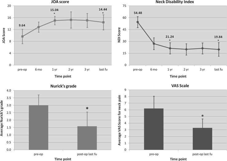
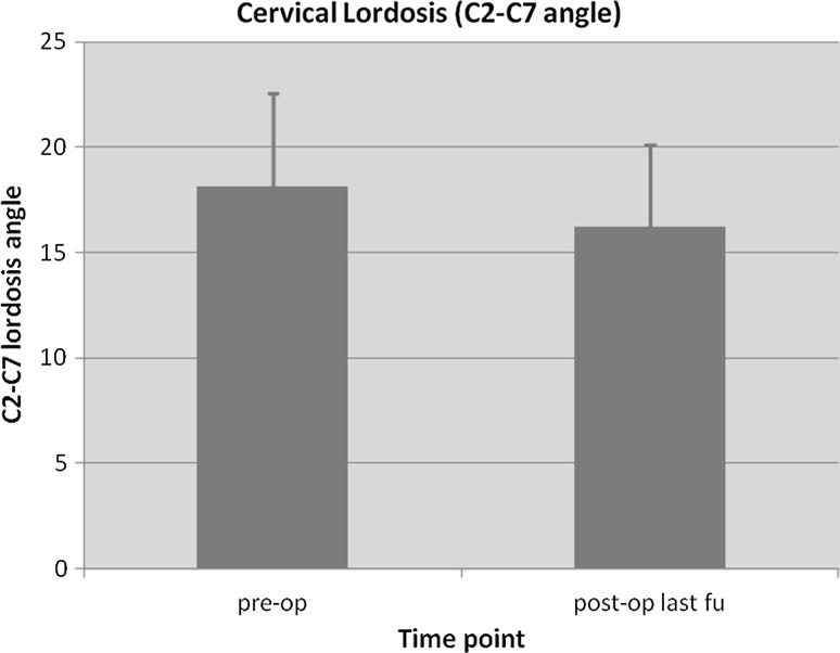
A series of 25 consecutive myelopathic patients were treated with a novel instrumented cervical French-door laminoplasty technique, whereby the enlarged posterior arch was held open with maxillofacial plates and screws. Patients had pre- and postoperative assessments with the Neck Disability Index (NDI), Japanese Orthopaedic Association (JOA) Score, Visual Analogue Score and radiographs. Minimum follow-up was 40 months, with regular interval assessments.
There were 18 males with a mean age of 45 years. The mean operative time was 145 min. The average hospital stay was 2.4 days and the mean follow-up was 56.5 months (40-72). All patients reported neurological improvements and there was a 35% improvement in NDI, and JOA score improved by 4.8 points. No postoperative hardware-related complications were noted and only one case of temporary C5 palsy, which completely resolved by the one-year follow-up.
Our data and clinical experience demonstrate that this hardware-augmented laminoplasty technique is safe and effective. We observed no hardware-related complications in our series. The use of readily available maxillofacial titanium miniplates and ease of surgical procedure means that this technique can be easily adopted into clinical practice.
Level IV.
用于治疗颈椎脊髓病的标准椎板切除术与继发性不稳定和后凸畸形相关,而椎板切除术联合融合术会使相邻节段有退变风险。单开门和双开门椎板成形术已被开发出来以克服这些局限性。最近,与植骨移位相关的并发症促使了器械辅助椎板成形术技术的发展。本研究的目的是回顾一种新开发的器械辅助法式开门椎板成形术治疗颈椎脊髓病的临床安全性和有效性。
连续25例脊髓病患者接受了一种新型的器械辅助颈椎法式开门椎板成形术,通过颌面钢板和螺钉将扩大的后弓撑开。患者术前和术后采用颈部功能障碍指数(NDI)、日本骨科协会(JOA)评分、视觉模拟评分和X线片进行评估。最短随访时间为40个月,并定期进行评估。
男性18例,平均年龄45岁。平均手术时间为145分钟。平均住院时间为2.4天,平均随访时间为56.5个月(40 - 72个月)。所有患者均报告神经功能有改善,NDI改善了35%,JOA评分提高了4.8分。未观察到术后与器械相关的并发症,仅1例出现暂时性C5麻痹,在1年随访时完全恢复。
我们的数据和临床经验表明,这种器械辅助椎板成形术技术是安全有效的。我们的系列研究中未观察到与器械相关的并发症。使用现成的颌面钛微型钢板以及手术操作简便意味着该技术可轻松应用于临床实践。
四级。