Tojima Michio, Ogata Naoshi, Nakahara Yasuo, Haga Nobuhiko
Department of Rehabilitation Medicine, the University of Tokyo, Tokyo, Japan; Department of Rehabilitation Medicine, the University of Tokyo Hospital, Japan; Faculty of Sport Sciences, Waseda University, Japan.
Department of Rehabilitation Medicine, the University of Tokyo Hospital, Japan; Department of Rehabilitation, Teikyo University, Japan.
J Hum Kinet. 2016 Apr 13;50:53-62. doi: 10.1515/hukin-2015-0141. eCollection 2016 Apr 1.
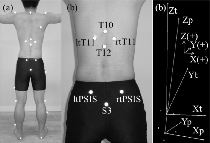
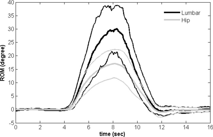
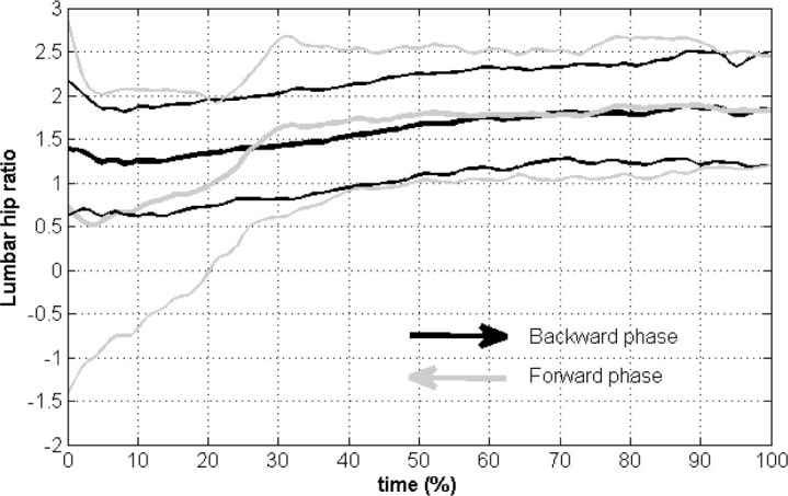
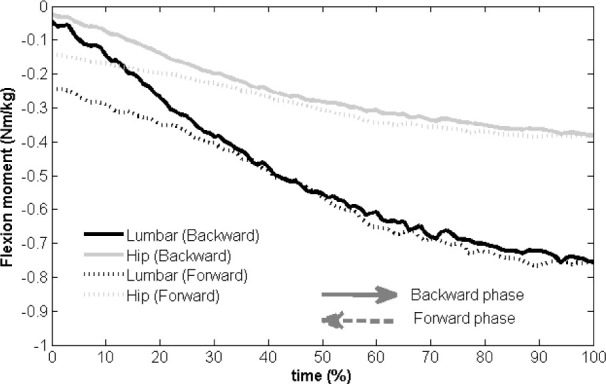
Hip-spine coordination, known as the lumbopelvic rhythm, can be expressed as the lumbar-hip ratio. The lumbopelvic rhythm and lumbar-hip ratio can be used to assess lower limb function. We clarified the lumbopelvic rhythm and lumbar-hip ratio during trunk extension. We established a novel set of marker positions for three-dimensional motion analysis to assess the lumbar spinal angle. The original markers were placed on both paravertebral muscle groups at the 11th thoracic spinous process level, the 10th and 12th thoracic spinous processes, and the pelvis. We measured angle data during trunk extension using three-dimensional motion analysis, and the data for eight healthy male subjects were categorized into backward and forward phases. The lumbar-hip ratio increased significantly from 1.2 to 1.9 (mean, 1.6) in the backward phase, indicating considerable movement of the lumbar spine compared with hip movement in the latter phase. In the forward phase, the ratio decreased significantly from 1.9 to 0.5 (mean, 1.5). After completion of 80% of the forward phase, the lumbar-hip ratio decreased to <1.0. The lumbopelvic rhythm for trunk extension was better expressed by a cubic or quadratic function than a linear function. According to a linear function, when the hip extends by 1°, lumbar spine extends by 1.9°. Therefore, lumbar spinal movement was greater than hip movement in the sagittal plane. The implication of the curved line would indicate lumbar extension instead of the limitation of hip extension.
髋部与脊柱的协调性,即腰骨盆节律,可用腰髋比来表示。腰骨盆节律和腰髋比可用于评估下肢功能。我们阐明了躯干伸展过程中的腰骨盆节律和腰髋比。我们建立了一套用于三维运动分析的新的标记点位置,以评估腰椎角度。原来的标记点放置在第11胸椎棘突水平的双侧椎旁肌群、第10和第12胸椎棘突以及骨盆处。我们使用三维运动分析测量了躯干伸展过程中的角度数据,并将八名健康男性受试者的数据分为后伸和前屈阶段。在后伸阶段,腰髋比从1.2显著增加到1.9(平均为1.6),这表明与后一阶段的髋部运动相比,腰椎有相当大的运动。在前屈阶段,该比值从1.9显著下降到0.5(平均为1.5)。在前屈阶段完成80%后,腰髋比降至<1.0。用三次函数或二次函数比用线性函数能更好地表示躯干伸展时的腰骨盆节律。根据线性函数,当髋部伸展1°时,腰椎伸展1.9°。因此,在矢状面上腰椎的运动大于髋部的运动。曲线的含义表明是腰椎伸展而不是髋部伸展受限。