Kling Sabine, Hammer Arthur, Conti Alain, Hafezi Farhad
Laboratory of Ocular Cell Biology, University of Geneva, Geneva, Switzerland ; Center for Applied Biotechnology and Molecular Medicine (CABMM), University of Zurich, Zurich, Switzerland.
Laboratory of Ocular Cell Biology, University of Geneva, Geneva, Switzerland.
Transl Vis Sci Technol. 2017 Jan 30;6(1):7. doi: 10.1167/tvst.6.1.7. eCollection 2017 Jan.
To morphologically, biochemically, and physiologically characterize corneal cross-linking with riboflavin and UV-A light (CXL) in a newly established in vivo murine model.
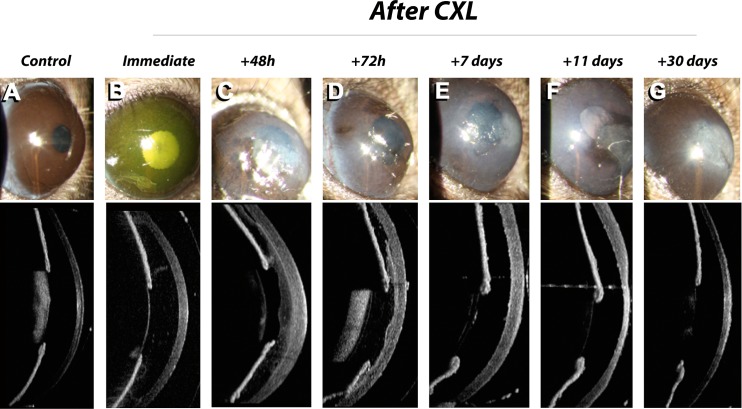
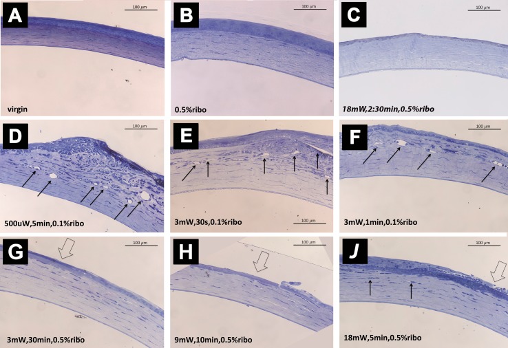
C57BL/6 wild-type mice ( = 67) were treated with various CXL protocols, with modification of the following parameters: total energy (fluence) used, duration of UV-A irradiation, continuous versus pulsed irradiation, and CXL under hypoxic conditions (contact lens). Corneas were evaluated biomicroscopically, histologically, and using optical coherence tomography. Conformational collagen changes were evaluated via changes in the speed of enzymatic digestion.
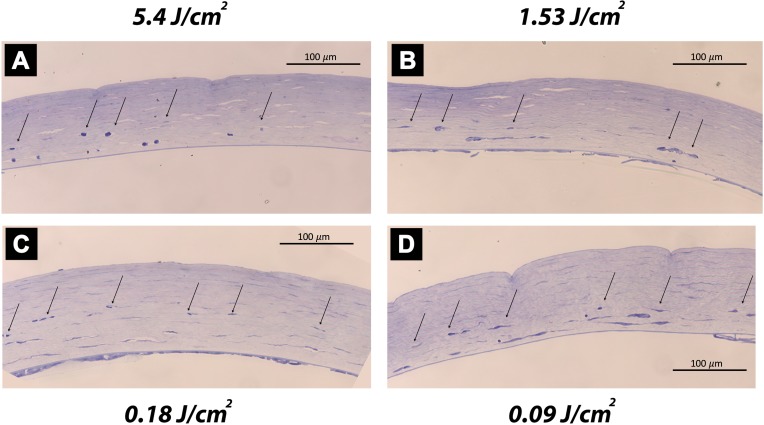
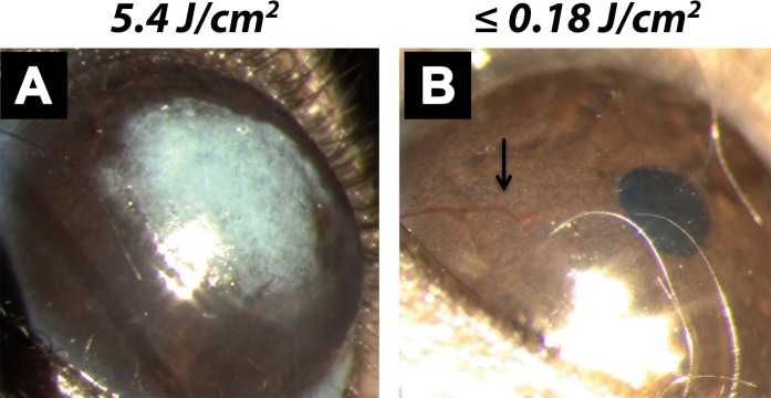
A fluence of 5.4 J/cm induced scar formation, while fluences of < 0.18 J/cm induced neovascularization. Fluences between 1.62 and 2.7 J/cm reduced epithelial thickness, but maintained a transparent cornea after 1 month. Pulsed UV irradiation inhibited neovascularization, but favored scar formation. Changes in the speed of enzymatic digestion suggest that CXL in mice, when compared to humans, requires less UV-A energy than the difference in corneal thickness between the species would suggest.
We demonstrated the in vivo response of very strong and very weak CXL and identified the best suited range of UV fluence in murine corneas. The presented murine CXL model may be helpful in future research addressing cellular and molecular pathways associated to CXL treatment.
Adverse tissue reactions following CXL treatment were observed, if the administered UV energy was out of the treatment window-raising concern about novel CXL treatment protocols that have not been previously validated in an experimental setting.
在新建立的体内小鼠模型中,对核黄素和紫外线A光角膜交联术(CXL)进行形态学、生物化学和生理学特征分析。
采用多种CXL方案处理67只C57BL/6野生型小鼠,改变以下参数:所用的总能量(通量)、紫外线A照射持续时间、连续照射与脉冲照射,以及低氧条件下(隐形眼镜)的CXL。通过生物显微镜检查、组织学检查和光学相干断层扫描对角膜进行评估。通过酶消化速度的变化评估胶原构象变化。
5.4 J/cm²的通量诱导瘢痕形成,而<0.18 J/cm²的通量诱导新生血管形成。1.62至2.7 J/cm²之间的通量可降低上皮厚度,但1个月后角膜仍保持透明。脉冲紫外线照射可抑制新生血管形成,但有利于瘢痕形成。酶消化速度的变化表明,与人类相比,小鼠的CXL所需的紫外线A能量比根据物种间角膜厚度差异所推测的要少。
我们展示了极强和极弱CXL的体内反应,并确定了小鼠角膜中最适合的紫外线通量范围。所提出的小鼠CXL模型可能有助于未来研究与CXL治疗相关的细胞和分子途径。
如果所给予的紫外线能量超出治疗窗口,CXL治疗后会观察到不良组织反应,这引发了对之前未在实验环境中验证的新型CXL治疗方案的担忧。