IMNC Laboratory, UMR 8165-CNRS/IN2P3, Paris-Saclay university, 91405 Orsay, France.
Neurosurgery Department, Sainte-Anne Hospital, France.
Sci Rep. 2017 Feb 2;7:41724. doi: 10.1038/srep41724.
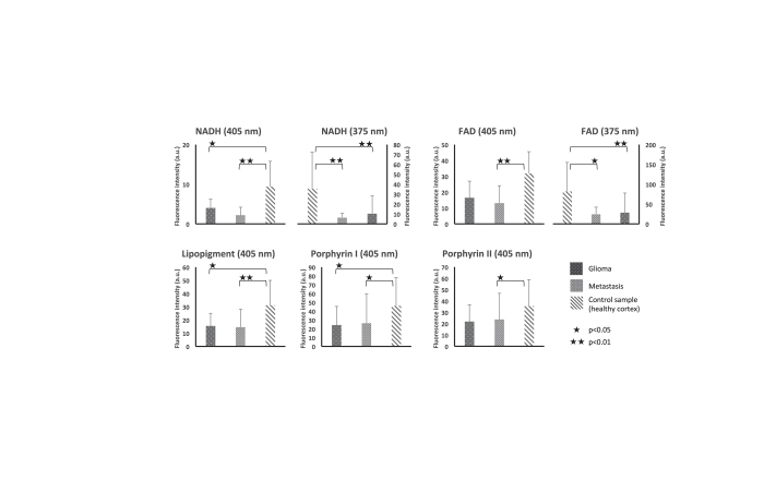
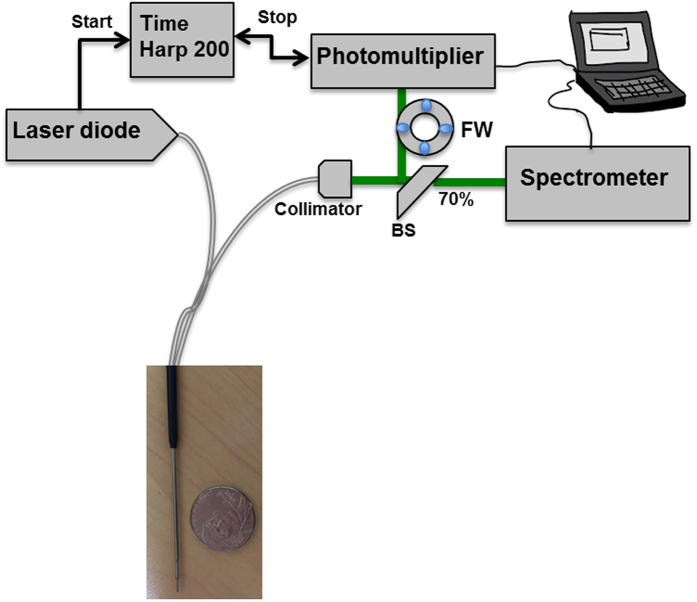
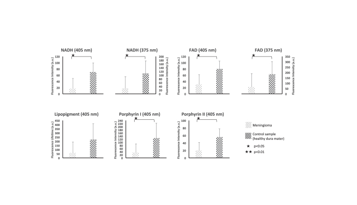
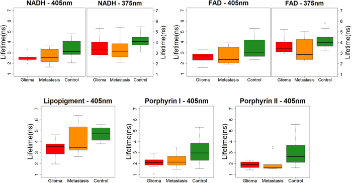
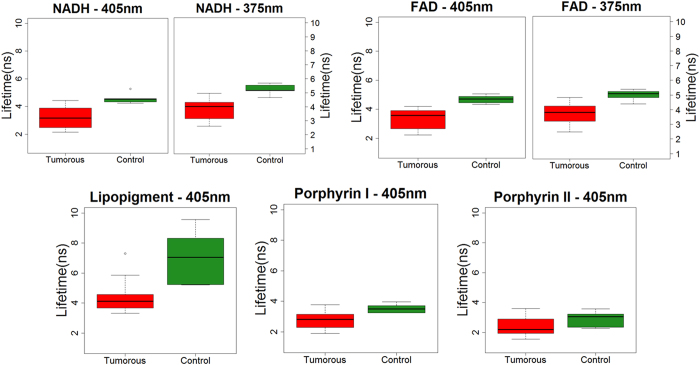
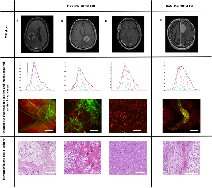
Delineating tumor margins as accurately as possible is of primordial importance in surgical oncology: extent of resection is associated with survival but respect of healthy surrounding tissue is necessary for preserved quality of life. The real-time analysis of the endogeneous fluorescence signal of brain tissues is a promising tool for defining margins of brain tumors. The present study aims to demonstrate the feasibility of multimodal optical analysis to discriminate fresh samples of gliomas, metastases and meningiomas from their appropriate controls. Tumor samples were studied on an optical fibered endoscope using spectral and fluorescence lifetime analysis and then on a multimodal set-up for acquiring spectral, one and two-photon fluorescence images, second harmonic generation signals and two-photon fluorescence lifetime datasets. The obtained data allowed us to differentiate healthy samples from tumor samples. These results confirmed the possible clinical relevance of this real-time multimodal optical analysis. This technique can be easily applied to neurosurgical procedures for a better delineation of surgical margins.
切除范围与生存率相关,但为了保持生活质量,必须保护周围健康组织。实时分析脑内组织的内源性荧光信号是定义脑肿瘤边界的一种很有前途的工具。本研究旨在证明多模态光学分析的可行性,以区分脑胶质瘤、转移瘤和脑膜瘤的新鲜样本与其相应对照的能力。使用光谱和荧光寿命分析,在光纤内窥镜上研究肿瘤样本,然后在多模态设备上获取光谱、单光子和双光子荧光图像、二次谐波产生信号和双光子荧光寿命数据集。获得的数据允许我们将健康样本与肿瘤样本区分开来。这些结果证实了这种实时多模态光学分析的可能临床相关性。该技术可轻松应用于神经外科手术,以更好地描绘手术边界。