Department of Chemistry and Chemical Biology, Harvard University, Cambridge, MA 02138, USA.
Sci Transl Med. 2013 Sep 4;5(201):201ra119. doi: 10.1126/scitranslmed.3005954.
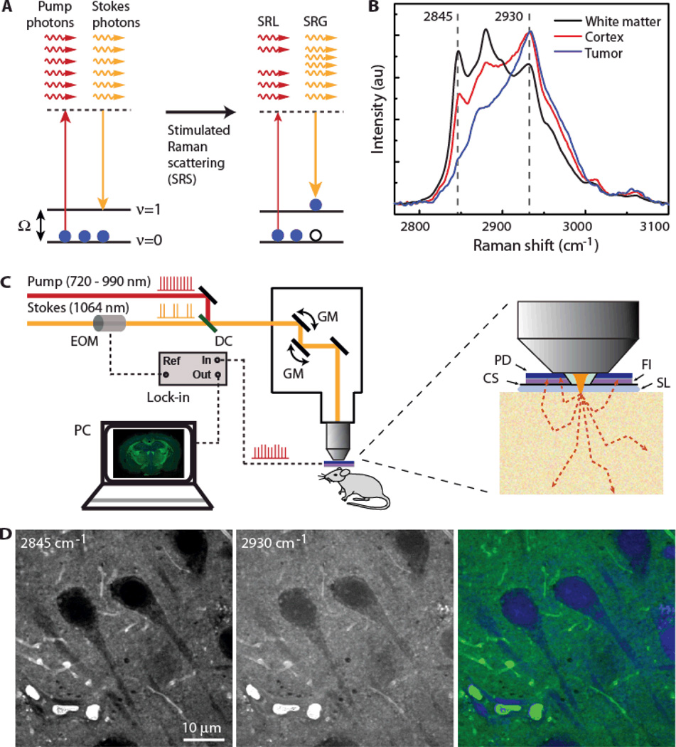
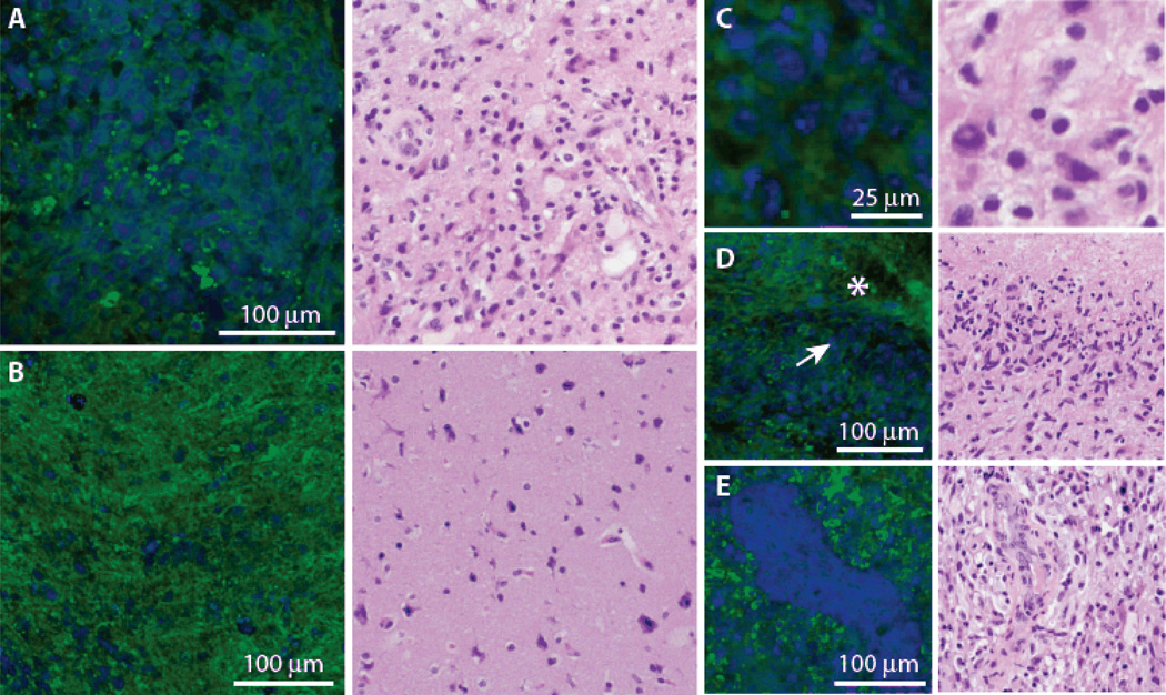
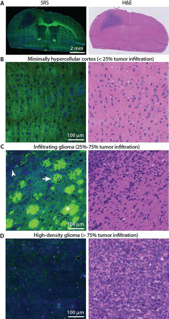
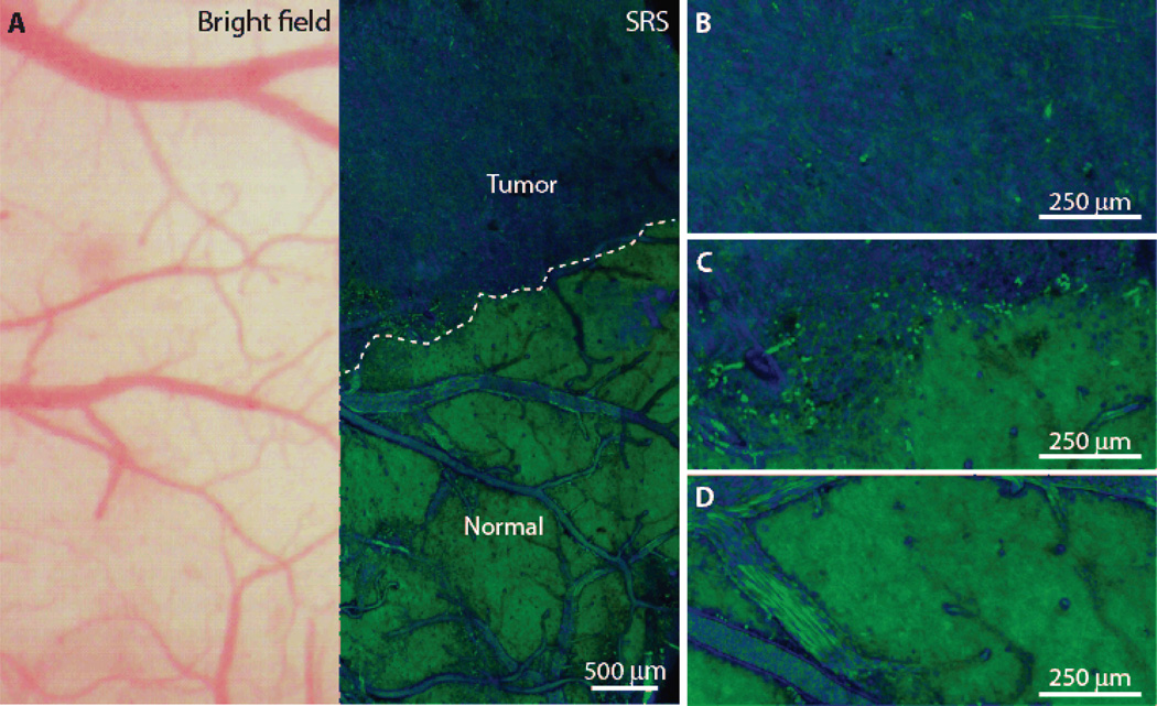
Surgery is an essential component in the treatment of brain tumors. However, delineating tumor from normal brain remains a major challenge. We describe the use of stimulated Raman scattering (SRS) microscopy for differentiating healthy human and mouse brain tissue from tumor-infiltrated brain based on histoarchitectural and biochemical differences. Unlike traditional histopathology, SRS is a label-free technique that can be rapidly performed in situ. SRS microscopy was able to differentiate tumor from nonneoplastic tissue in an infiltrative human glioblastoma xenograft mouse model based on their different Raman spectra. We further demonstrated a correlation between SRS and hematoxylin and eosin microscopy for detection of glioma infiltration (κ = 0.98). Finally, we applied SRS microscopy in vivo in mice during surgery to reveal tumor margins that were undetectable under standard operative conditions. By providing rapid intraoperative assessment of brain tissue, SRS microscopy may ultimately improve the safety and accuracy of surgeries where tumor boundaries are visually indistinct.
手术是治疗脑肿瘤的重要组成部分。然而,将肿瘤与正常脑组织区分开来仍然是一个主要挑战。我们描述了使用受激拉曼散射(SRS)显微镜根据组织形态学和生化差异来区分健康的人和鼠脑组织与浸润性肿瘤脑组织。与传统的组织病理学不同,SRS 是一种无需标记的技术,可以在原位快速进行。SRS 显微镜能够根据不同的拉曼光谱区分浸润性人胶质母细胞瘤异种移植小鼠模型中的肿瘤与非肿瘤组织。我们进一步证明了 SRS 与苏木精和伊红显微镜检测胶质瘤浸润之间的相关性(κ=0.98)。最后,我们在手术过程中在小鼠体内应用 SRS 显微镜来揭示在标准手术条件下无法检测到的肿瘤边界。通过提供对脑组织的快速术中评估,SRS 显微镜可能最终提高在肿瘤边界肉眼不可分辨的手术中的安全性和准确性。