Lewis Aaran T, Gaifulina Riana, Isabelle Martin, Dorney Jennifer, Woods Mae L, Lloyd Gavin R, Lau Katherine, Rodriguez-Justo Manuel, Kendall Catherine, Stone Nicholas, Thomas Geraint M
Department of Cell and Developmental Biology University College London London UK.
Biophotonics Research Unit Gloucester Royal Hospitals NHS Foundation Trust Gloucestershire UK.
J Raman Spectrosc. 2017 Jan;48(1):119-125. doi: 10.1002/jrs.4980. Epub 2016 Jul 29.
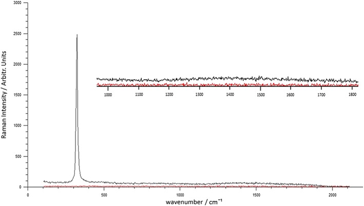
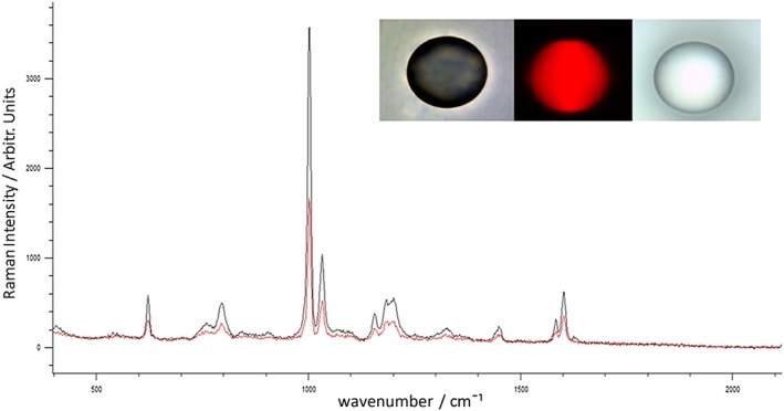
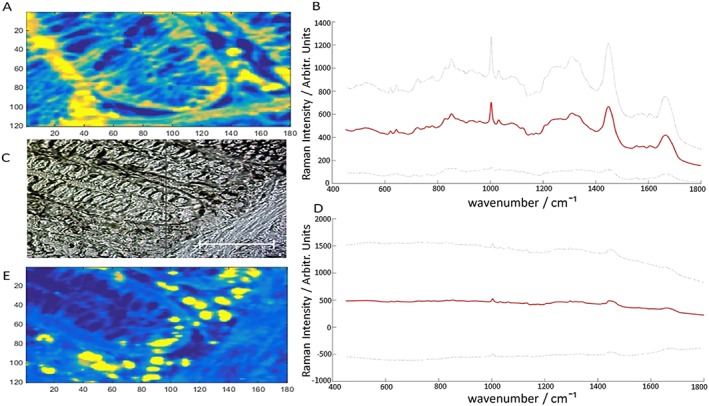
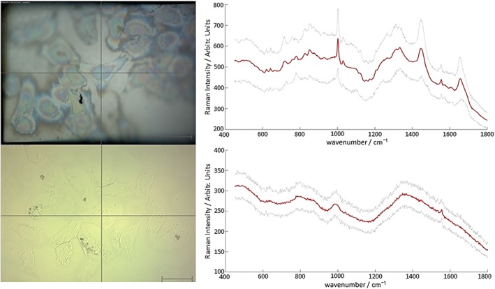
Raman spectroscopy (RS) is a powerful technique that permits the non-destructive chemical analysis of cells and tissues without the need for expensive and complex sample preparation. To date, samples have been routinely mounted onto calcium fluoride (CaF) as this material possesses the desired mechanical and optical properties for analysis, but CaF is both expensive and brittle and this prevents the technique from being routinely adopted. Furthermore, Raman scattering is a weak phenomenon and CaF provides no means of increasing signal. For RS to be widely adopted, particularly in the clinical field, it is crucial that spectroscopists identify an alternative, low-cost substrate capable of providing high spectral signal to noise ratios with good spatial resolution. Results show that these desired properties are attainable when using mirrored stainless steel as a Raman substrate. When compared with CaF, data show that stainless steel has a low background signal and provides an average signal increase of 1.43 times during tissue analysis and 1.64 times when analyzing cells. This result is attributed to a double-pass of the laser beam through the sample where the photons from the source laser and the forward scattered Raman signal are backreflected and retroreflected from the mirrored steel surface and focused towards collection optics. The spatial resolution on stainless steel is at least comparable to that on CaF and it is not compromised by the reflection of the laser. Steel is a fraction of the cost of CaF and the reflection and focusing of photons improve signal to noise ratios permitting more rapid mapping. The low cost of steel coupled with its Raman signal increasing properties and robust durability indicates that steel is an ideal substrate for biological and clinical RS as it possesses key advantages over routinely used CaF. © 2016 The Authors. Published by John Wiley & Sons Ltd.
拉曼光谱(RS)是一种强大的技术,它能够对细胞和组织进行无损化学分析,而无需进行昂贵且复杂的样品制备。迄今为止,样品通常被安装在氟化钙(CaF)上,因为这种材料具有分析所需的机械和光学特性,但CaF既昂贵又易碎,这使得该技术无法被常规采用。此外,拉曼散射是一种微弱的现象,CaF无法提高信号。为了使RS被广泛采用,特别是在临床领域,光谱学家确定一种能够提供高光谱信噪比且具有良好空间分辨率的替代低成本基质至关重要。结果表明,使用镜面不锈钢作为拉曼基质时可以实现这些所需的特性。与CaF相比,数据显示不锈钢具有低背景信号,在组织分析期间平均信号增加1.43倍,分析细胞时增加1.64倍。这一结果归因于激光束两次穿过样品,其中来自源激光的光子和向前散射的拉曼信号从镜面钢表面被反向反射和后向反射,并聚焦到收集光学器件上。不锈钢上的空间分辨率至少与CaF上的相当,并且不会因激光反射而受到影响。钢的成本只是CaF的一小部分,光子的反射和聚焦提高了信噪比,从而允许更快速地绘制图谱。钢的低成本及其拉曼信号增强特性和强大的耐用性表明,钢是生物和临床RS的理想基质,因为它比常规使用的CaF具有关键优势。© 2016作者。由约翰·威利父子有限公司出版。