Arnold Douglas L, Calabresi Peter A, Kieseier Bernd C, Liu Shifang, You Xiaojun, Fiore Damian, Hung Serena
Montreal Neurological Institute, McGill University, Montreal, QC, Canada.
NeuroRx Research, Montreal, QC, Canada.
BMC Neurol. 2017 Feb 10;17(1):29. doi: 10.1186/s12883-017-0799-0.
Subcutaneous peginterferon beta-1a has previously been shown to reduce the number of T2-hyperintense and gadolinium-enhancing (Gd+) lesions over 2 years in patients with relapsing-remitting multiple sclerosis (RRMS), and to reduce T1-hypointense lesion formation and the proportion of patients showing evidence of disease activity, based on both clinical and radiological measures, compared with placebo over 1 year of treatment. The objectives of the current analyses were to evaluate T1 lesions and other magnetic resonance imaging (MRI) measures, including whole brain volume and magnetization transfer ratio (MTR) of normal appearing brain tissue (NABT), and the proportions of patients with no evidence of disease activity (NEDA), over 2 years.
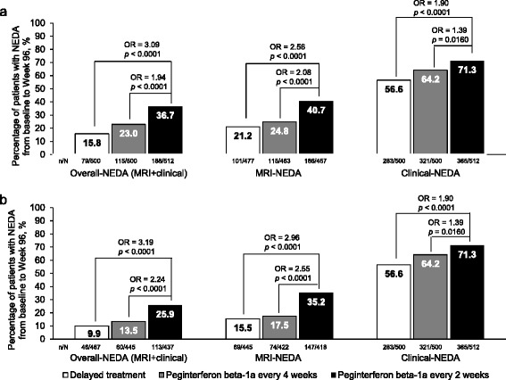
Patients enrolled in the ADVANCE study received continuous peginterferon beta-1a every 2 or 4 weeks for 2 years, or delayed treatment (placebo in Year 1; peginterferon beta-1a every 2 or 4 weeks in Year 2). MRI scans were performed at baseline and Weeks 24, 48, and 96. Proportions of patients with NEDA were calculated based on radiological criteria (absence of Gd + and new/newly-enlarging T2 lesions) and clinical criteria (no relapse or confirmed disability progression) separately and overall.
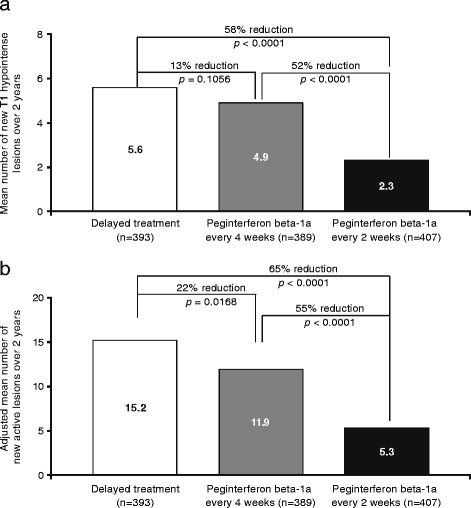
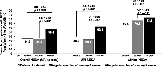
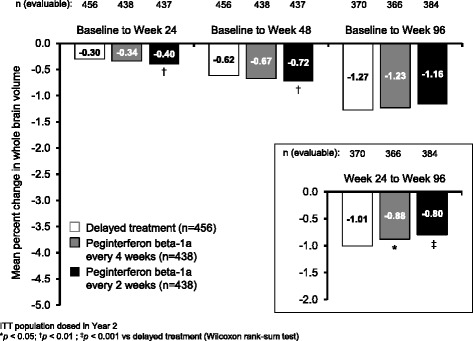
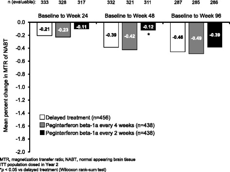
Peginterferon beta-1a every 2 weeks significantly reduced the number and volume of T1-hypointense lesions compared with delayed treatment over 2 years. Changes in whole brain volume and MTR of NABT were suggestive of pseudoatrophy during the first 6 months of peginterferon beta-1a treatment, which subsequently began to resolve. Significantly more patients in the peginterferon beta-1a every 2 weeks group compared with the delayed treatment group met MRI-NEDA criteria (41% vs 21%; odds ratio [OR] 2.56; p < 0.0001), clinical-NEDA criteria (71% vs 57%; OR 1.90; p < 0.0001) and achieved overall-NEDA (37% vs 16%; OR 3.09; p < 0.0001).
Peginterferon beta-1a provides significant improvements in MRI measures and offers patients a good chance of remaining free from evidence of MRI, clinical and overall disease activity over a sustained 2-year period.
ClinicalTrials.gov: NCT00906399 ; Registered on: May 20, 2009.
先前的研究表明,皮下注射聚乙二醇化干扰素β-1a可使复发缓解型多发性硬化症(RRMS)患者在2年内T2高信号和钆增强(Gd+)病灶数量减少,并且与治疗1年的安慰剂相比,基于临床和影像学测量,皮下注射聚乙二醇化干扰素β-1a还可减少T1低信号病灶形成以及出现疾病活动证据的患者比例。本次分析的目的是评估2年内的T1病灶及其他磁共振成像(MRI)指标,包括全脑体积和正常脑组织(NABT)的磁化传递率(MTR),以及无疾病活动证据(NEDA)患者的比例。
参加ADVANCE研究的患者每2周或4周持续接受聚乙二醇化干扰素β-1a治疗2年,或延迟治疗(第1年使用安慰剂;第2年每2周或4周使用聚乙二醇化干扰素β-1a)。在基线、第24周、第48周和第96周进行MRI扫描。分别根据影像学标准(无Gd+和新出现/新增大的T2病灶)和临床标准(无复发或确诊的残疾进展)以及总体标准计算NEDA患者的比例。
与延迟治疗相比,每2周一次皮下注射聚乙二醇化干扰素β-1a在2年内显著减少了T1低信号病灶的数量和体积。NABT的全脑体积和MTR变化提示在皮下注射聚乙二醇化干扰素β-1a治疗的前6个月出现了假性萎缩,随后开始缓解。与延迟治疗组相比,每2周一次皮下注射聚乙二醇化干扰素β-1a组达到MRI-NEDA标准(41%对21%;优势比[OR]2.56;p<0.0001)、临床-NEDA标准(71%对57%;OR 1.90;p<0.0001)和总体-NEDA标准(37%对16%;OR 3.09;p<0.0001)的患者明显更多。
聚乙二醇化干扰素β-1a在MRI指标方面有显著改善,并为患者提供了在持续2年期间无MRI、临床和总体疾病活动证据的良好机会。
ClinicalTrials.gov:NCT00906399;注册时间:2009年5月20日。