Curry Joseph, Johnson Jennifer, Tassone Patrick, Vidal Marina Domingo, Menezes Diana Whitaker, Sprandio John, Mollaee Mehri, Cotzia Paolo, Birbe Ruth, Lin Zhao, Gill Kurren, Duddy Elizabeth, Zhan Tingting, Leiby Benjamin, Reyzer Michelle, Cognetti David, Luginbuhl Adam, Tuluc Madalina, Martinez-Outschoorn Ubaldo
Department of Otolaryngology-Head and Neck Surgery, Philadelphia, Pennsylvania, U.S.A.
Department of Medical Oncology, Philadelphia, Pennsylvania, U.S.A.
Laryngoscope. 2017 Aug;127(8):1808-1815. doi: 10.1002/lary.26489. Epub 2017 Feb 10.
The tumor microenvironment frequently displays abnormal cellular metabolism, which contributes to aggressive behavior. Metformin inhibits mitochondrial oxidative phosphorylation, altering metabolism. Though the mechanism is unclear, epidemiologic studies show an association between metformin use and improved outcomes in head and neck squamous cell carcinoma (HNSCC). We sought to determine if metformin alters metabolism and apoptosis in HNSCC tumors.
Window of opportunity trial of metformin between diagnostic biopsy and resection. Participants were patients with newly diagnosed HNSCC. Fifty patients were enrolled, and 39 completed a full-treatment course. Metformin was titrated to standard diabetic dose (2,000 mg/day) for a course of 9 or more days prior to surgery.
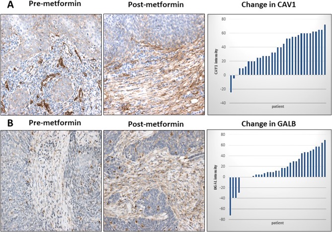
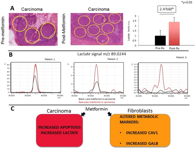
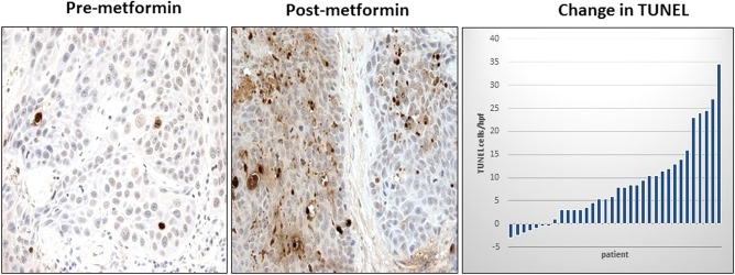
Immunohistochemistry (IHC) for the metabolic markers caveolin-1 (CAV1), B-galactosidase (GALB), and monocarboxylate transporter 4 (MCT4), as well as the Terminal deoxynucleotidyl transferase dUTP nick end labeling (TUNEL) apoptosis assay and Ki-67 IHC, were performed in pre- and postmetformin specimens. Exploratory mass spectroscopy imaging (MSI) to assess lactate levels also was performed in three subjects.
Metformin was well tolerated. The average treatment course was 13.6 days. Posttreatment specimens showed a significant increase in stromal CAV1 (P < 0.001) and GALB (P < 0.005), as well as tumor cell apoptosis by TUNEL assay (P < 0.001). There was no significant change in stromal MCT4 expression or proliferation measured by Ki67. Lactate levels in carcinoma cells were increased 2.4-fold postmetformin (P < 0.05), as measured by MSI.
Metformin increases markers of reduced catabolism and increases senescence in stromal cells as well as carcinoma cell apoptosis. This study demonstrates that metformin modulates metabolism in the HNSCC microenvironment.
肿瘤微环境常表现出异常的细胞代谢,这会导致侵袭性生物学行为。二甲双胍可抑制线粒体氧化磷酸化,从而改变代谢。尽管其机制尚不清楚,但流行病学研究表明,使用二甲双胍与头颈部鳞状细胞癌(HNSCC)预后改善之间存在关联。我们试图确定二甲双胍是否会改变HNSCC肿瘤的代谢和凋亡。
在诊断性活检和手术切除之间进行二甲双胍的机会性试验。参与者为新诊断的HNSCC患者。共招募了50名患者,其中39名完成了整个治疗疗程。在手术前,将二甲双胍滴定至标准糖尿病剂量(2000毫克/天),疗程为9天或更长时间。
对二甲双胍治疗前后的标本进行免疫组织化学(IHC)检测,检测代谢标志物小窝蛋白-1(CAV1)、β-半乳糖苷酶(GALB)和单羧酸转运蛋白4(MCT4),以及末端脱氧核苷酸转移酶dUTP缺口末端标记(TUNEL)凋亡检测和Ki-67 IHC。还对三名受试者进行了探索性质谱成像(MSI)以评估乳酸水平。
二甲双胍耐受性良好。平均治疗疗程为13.6天。治疗后的标本显示,基质CAV1(P < 0.001)和GALB(P < 0.005)显著增加,通过TUNEL检测发现肿瘤细胞凋亡也显著增加(P < 0.001)。通过Ki67检测,基质MCT4表达或增殖无显著变化。通过MSI检测,二甲双胍治疗后癌细胞中的乳酸水平增加了2.4倍(P < 0.05)。
二甲双胍增加了分解代谢降低的标志物,并增加了基质细胞的衰老以及癌细胞的凋亡。本研究表明,二甲双胍可调节HNSCC微环境中的代谢。
4。《喉镜》,2017年,第127卷,第1808 - 1815页。