Kao Yu-Chieh Jill, Oyarzabal Esteban A, Zhang Hua, Faber James E, Shih Yen-Yu Ian
From the Department of Neurology (Y.-C.J.K., E.A.O.), Biomedical Research Imaging Center (Y.-C.J.K., E.A.O., Y.-Y.I.S.), Neurobiology Curriculum (E.A.O., J.E.F.), Department of Cell Biology and Physiology (H.Z., J.E.F.), McAllister Heart Institute (H.Z., J.E.F., Y.-Y.I.S.), and Department of Biomedical Engineering (Y.-Y.I.S.), University of North Carolina, Chapel Hill; and Translational Imaging Research Center (Y.-C.J.K.) and Department of Radiology, School of Medicine (Y.-C.J.K.), College of Medicine, Taipei Medical University, Taiwan.
Stroke. 2017 Mar;48(3):754-761. doi: 10.1161/STROKEAHA.116.015878. Epub 2017 Feb 10.
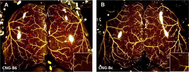
No studies have determined the effect of differences in pial collateral extent (number and diameter), independent of differences in environmental factors and unknown genetic factors, on severity of stroke. We examined ischemic tissue evolution during acute stroke, as measured by magnetic resonance imaging and histology, by comparing 2 congenic mouse strains with otherwise identical genetic backgrounds but with different alleles of the () genetic locus. We also optimized magnetic resonance perfusion and diffusion-deficit thresholds by using histological measures of ischemic tissue.
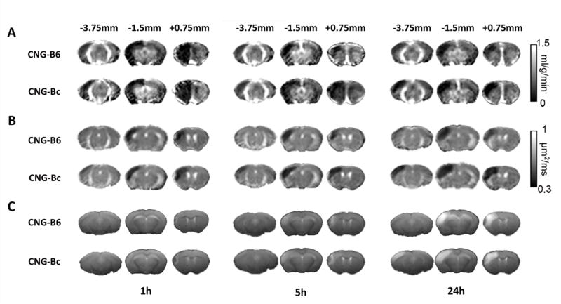
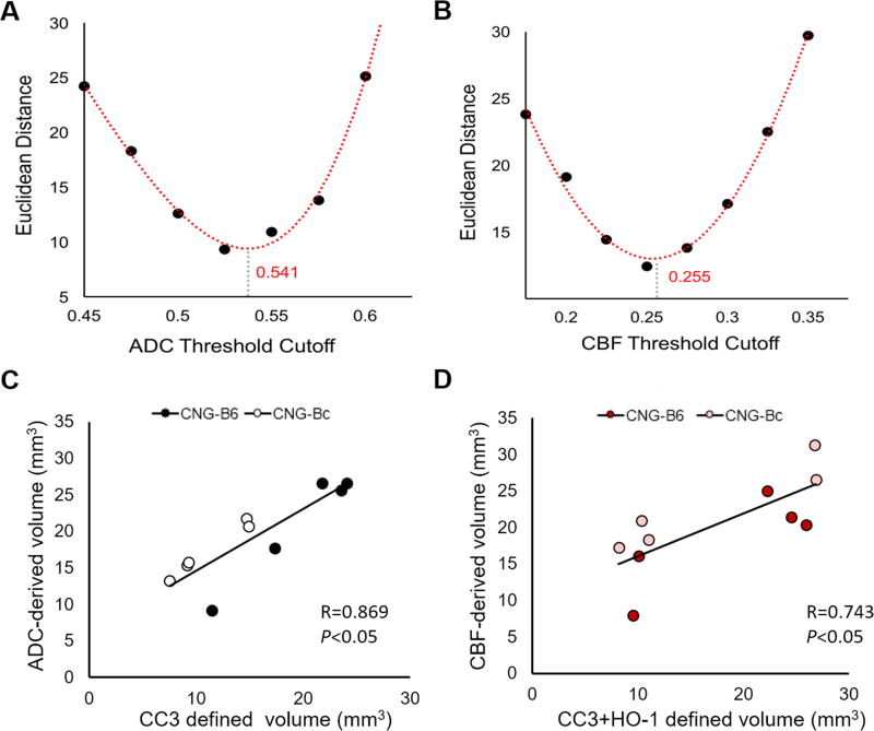
Perfusion, diffusion, and T-weighted magnetic resonance imaging were performed on collateral-poor (congenic-Bc) and collateral-rich (congenic-B6) mice at 1, 5, and 24 hours after permanent middle cerebral artery occlusion. Magnetic resonance imaging-derived penumbra and ischemic core volumes were confirmed by histology in a subset of mice at 5 and 24 hours after permanent middle cerebral artery occlusion.
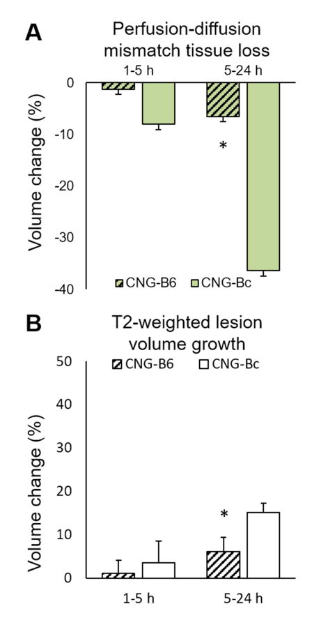
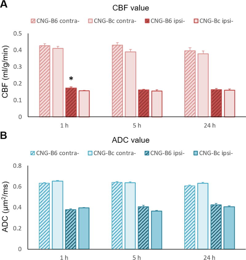
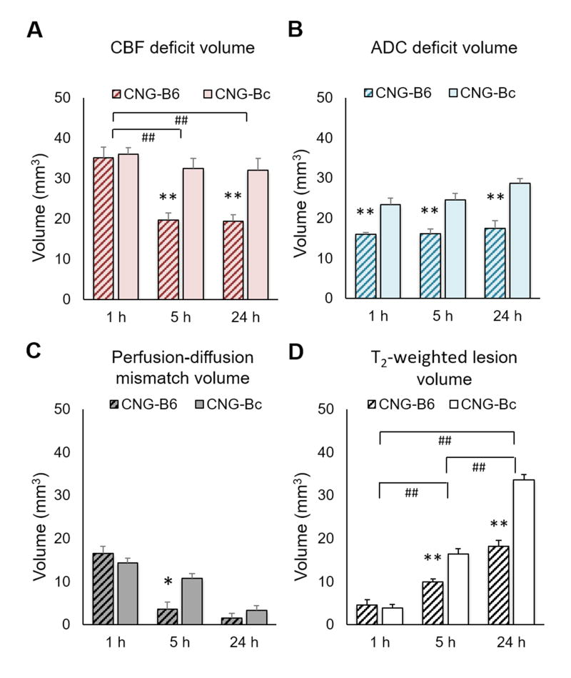
Although perfusion-deficit volumes were similar between strains 1 hour after permanent middle cerebral artery occlusion, diffusion-deficit volumes were 32% smaller in collateral-rich mice. At 5 hours, collateral-rich mice had markedly restored perfusion patterns showing reduced perfusion-deficit volumes, smaller infarct volumes, and smaller perfusion-diffusion mismatch volumes compared with the collateral-poor mice (<0.05). At 24 hours, collateral-rich mice had 45% smaller T-weighted lesion volumes (<0.005) than collateral-poor mice, with no difference in perfusion-diffusion mismatch volumes because of penumbral death occurring 5 to 24 hours after permanent middle cerebral artery occlusion in collateral-poor mice.
Variation in collateral extent significantly alters infarct volume expansion, transiently affects perfusion and diffusion magnetic resonance imaging signatures, and impacts salvage of ischemic penumbra after stroke onset.
尚无研究确定软脑膜侧支程度(数量和直径)的差异(独立于环境因素和未知遗传因素的差异)对卒中严重程度的影响。我们通过比较两种具有相同遗传背景但()基因座等位基因不同的近交系小鼠,研究了急性卒中期间缺血组织的演变,采用磁共振成像和组织学方法进行测量。我们还通过使用缺血组织的组织学测量方法优化了磁共振灌注和扩散缺损阈值。
在永久性大脑中动脉闭塞后1、5和24小时,对侧支少(近交系-Bc)和侧支丰富(近交系-B6)的小鼠进行灌注、扩散和T加权磁共振成像。在永久性大脑中动脉闭塞后5和24小时,通过组织学方法在一部分小鼠中确认了磁共振成像得出的半暗带和缺血核心体积。
尽管在永久性大脑中动脉闭塞后1小时,各品系之间的灌注缺损体积相似,但侧支丰富的小鼠的扩散缺损体积小32%。在5小时时,与侧支少的小鼠相比,侧支丰富的小鼠灌注模式明显恢复,灌注缺损体积减小,梗死体积减小,灌注-扩散不匹配体积减小(<0.05)。在24小时时,侧支丰富的小鼠的T加权病变体积比侧支少的小鼠小45%(<0.005),由于侧支少的小鼠在永久性大脑中动脉闭塞后5至24小时发生半暗带死亡,灌注-扩散不匹配体积没有差异。
侧支程度的变化显著改变梗死体积的扩大,短暂影响灌注和扩散磁共振成像特征,并影响卒中发作后缺血半暗带的挽救。