Grah Joana Sarah, Harrington Jennifer Alison, Koh Siang Boon, Pike Jeremy Andrew, Schreiner Alexander, Burger Martin, Schönlieb Carola-Bibiane, Reichelt Stefanie
University of Cambridge, Department of Applied Mathematics and Theoretical Physics, Centre for Mathematical Sciences, Wilberforce Road, Cambridge CB3 0WA, United Kingdom.
University of Cambridge, Cancer Research UK Cambridge Institute, Li Ka Shing Centre, Robinson Way, Cambridge CB2 0RE, United Kingdom.
Methods. 2017 Feb 15;115:91-99. doi: 10.1016/j.ymeth.2017.02.001. Epub 2017 Feb 9.
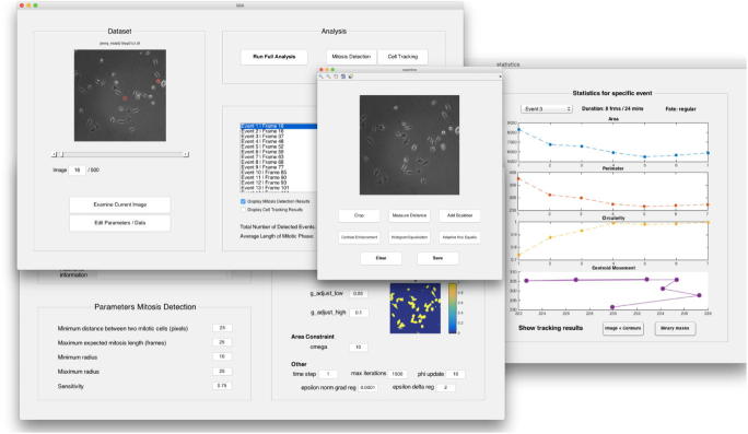
In this paper we propose a workflow to detect and track mitotic cells in time-lapse microscopy image sequences. In order to avoid the requirement for cell lines expressing fluorescent markers and the associated phototoxicity, phase contrast microscopy is often preferred over fluorescence microscopy in live-cell imaging. However, common specific image characteristics complicate image processing and impede use of standard methods. Nevertheless, automated analysis is desirable due to manual analysis being subjective, biased and extremely time-consuming for large data sets. Here, we present the following workflow based on mathematical imaging methods. In the first step, mitosis detection is performed by means of the circular Hough transform. The obtained circular contour subsequently serves as an initialisation for the tracking algorithm based on variational methods. It is sub-divided into two parts: in order to determine the beginning of the whole mitosis cycle, a backwards tracking procedure is performed. After that, the cell is tracked forwards in time until the end of mitosis. As a result, the average of mitosis duration and ratios of different cell fates (cell death, no division, division into two or more daughter cells) can be measured and statistics on cell morphologies can be obtained. All of the tools are featured in the user-friendly MATLAB®Graphical User Interface MitosisAnalyser.
在本文中,我们提出了一种用于在延时显微镜图像序列中检测和跟踪有丝分裂细胞的工作流程。为了避免对表达荧光标记的细胞系的需求以及相关的光毒性,在活细胞成像中,相衬显微镜通常比荧光显微镜更受青睐。然而,常见的特定图像特征使图像处理变得复杂,并阻碍了标准方法的使用。尽管如此,由于手动分析具有主观性、偏差性且对于大数据集极其耗时,因此自动化分析是很有必要的。在此,我们基于数学成像方法提出以下工作流程。第一步,通过圆形霍夫变换进行有丝分裂检测。所获得的圆形轮廓随后用作基于变分方法的跟踪算法的初始化。它分为两个部分:为了确定整个有丝分裂周期的开始,执行向后跟踪程序。之后,对细胞进行及时向前跟踪,直到有丝分裂结束。结果,可以测量有丝分裂持续时间的平均值以及不同细胞命运(细胞死亡、不分裂、分裂为两个或更多子细胞)的比例,并获得细胞形态的统计数据。所有工具都集成在用户友好的MATLAB®图形用户界面MitosisAnalyser中。