Furdová Alena, Sramka Miron, Thurzo Andrej, Furdová Adriana
Department of Ophthalmology, Faculty of Medicine, Comenius University.
Department of Stereotactic Radiosurgery, St Elisabeth Cancer Inst and St Elisabeth University College of Health and Social Work.
Clin Ophthalmol. 2017 Jan 31;11:267-271. doi: 10.2147/OPTH.S123640. eCollection 2017.
The objective of this study was to determine the use of 3D printed model of an eye with intraocular tumor for linear accelerator-based stereotactic radiosurgery.
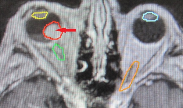
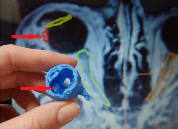
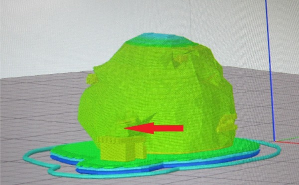
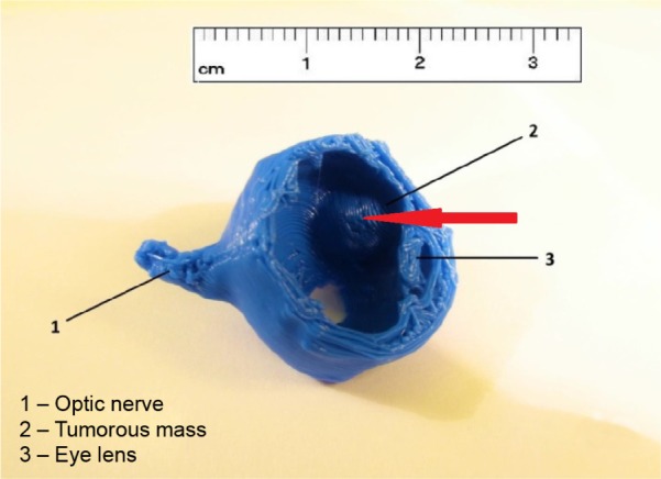
The software for segmentation (3D Slicer) created virtual 3D model of eye globe with tumorous mass based on tissue density from computed tomography and magnetic resonance imaging data. A virtual model was then processed in the slicing software (Simplify3D) and printed on 3D printer using fused deposition modeling technology. The material that was used for printing was polylactic acid.
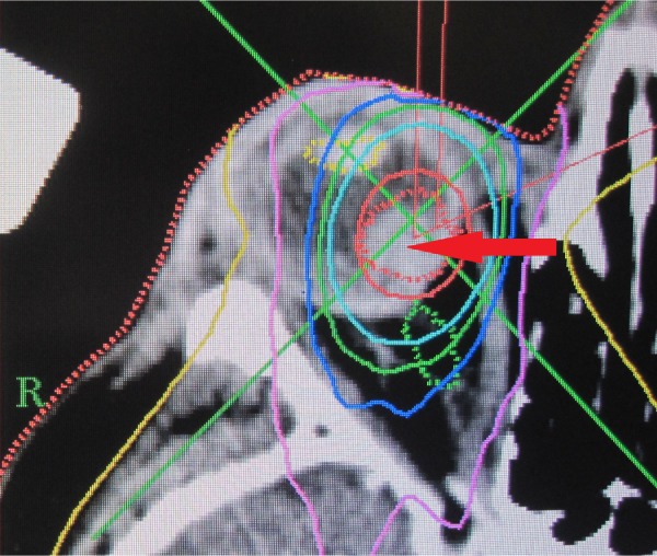
In 2015, stereotactic planning scheme was optimized with the help of 3D printed model of the patient's eye with intraocular tumor. In the period 2001-2015, a group of 150 patients with uveal melanoma (139 choroidal melanoma and 11 ciliary body melanoma) were treated. The median tumor volume was 0.5 cm (0.2-1.6 cm). The radiation dose was 35.0 Gy by 99% of dose volume histogram.
The 3D printed model of eye with tumor was helpful in planning the process to achieve the optimal scheme for irradiation which requires high accuracy of defining the targeted tumor mass and critical structures.
本研究的目的是确定使用带有眼内肿瘤的眼睛的3D打印模型进行基于直线加速器的立体定向放射外科手术。
分割软件(3D Slicer)根据计算机断层扫描和磁共振成像数据中的组织密度创建带有肿瘤块的眼球虚拟3D模型。然后在切片软件(Simplify3D)中对虚拟模型进行处理,并使用熔融沉积建模技术在3D打印机上打印。用于打印的材料是聚乳酸。
2015年,借助带有眼内肿瘤的患者眼睛的3D打印模型优化了立体定向计划方案。在2001年至2015年期间,对一组150例葡萄膜黑色素瘤患者(139例脉络膜黑色素瘤和11例睫状体黑色素瘤)进行了治疗。肿瘤体积中位数为0.5 cm(0.2 - 1.6 cm)。通过剂量体积直方图的99%,放射剂量为35.0 Gy。
带有肿瘤的眼睛的3D打印模型有助于规划实现最佳照射方案的过程,该方案需要高精度地定义目标肿瘤块和关键结构。