Department of Ophthalmology, Faculty of Medicine, Hospital Ruzinov, Comenius University Bratislava, Ruzinovska 6, 826 06, Bratislava, Slovak Republic.
Department of Information Systems, Faculty of Management, Comenius University Bratislava, Odbojarov 10, 820 05, Bratislava, Slovak Republic.
BMC Ophthalmol. 2022 Aug 5;22(1):333. doi: 10.1186/s12886-022-02558-w.
Stereotactic irradiation is one of the treatment modalities for intraocular uveal melanoma. The study's purpose was to describe the background of stereotactic one-day session radiosurgery, how the comparison in the difference between the tumor volume measured values from the magnetic resonance imaging (MRI) method and the ultrasound method was related to it, and which method was more precise to be used for tumor regression after irradiation.
The group of 147 patients with choroidal melanoma was treated by stereotactic irradiation on the linear accelerator with a single dose of 35.0 Gy. During the standard treatment process the uveal melanoma volumes, needed for dose calculation, were obtained using MRI from the individual stereotactic planning scheme and by ultrasound from the ultrasound device. All volumes were statistically compared using the paired t-test, and for the visualization purpose, the Bland-Altman plot was used.
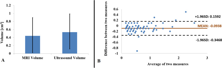
In the group of patients, it was 70 (47.6%) males and 77 (52.4%) females. The tumor volume median was from MRI equal to 0.44 cm and from ultrasound equal to 0.53 cm. The difference between the ultrasound and the MRI volume measured values was statistically significant. However, the Bland-Altman plot clearly documents that the two methods are in agreement and can be used interchangeably. In most of the cases, the measured values of the ultrasound-calculated volume achieved slightly higher measured values.
The calculation of the intraocular uveal tumor volume is a crucial part of the stereotactic irradiation treatment. The ultrasound volume measured values were in most of the cases higher than the measured values from the MRI. Although the methods are comparable and can be used interchangeably, we are recommending using the more precise MRI method not only during the treatment but also on later regular medical checks of tumor regression or progression.
立体定向放射治疗是治疗眼内葡萄膜黑色素瘤的一种方法。本研究旨在描述立体定向一日疗程放射外科的背景,探讨肿瘤体积测量值的差异与 MRI 方法和超声方法之间的关系,以及哪种方法更精确,更适用于肿瘤照射后的消退。
147 例脉络膜黑色素瘤患者在直线加速器上进行立体定向照射,单次剂量为 35.0Gy。在标准治疗过程中,使用个体立体定向计划方案中的 MRI 和超声设备获得用于剂量计算的葡萄膜黑色素瘤体积。所有体积均采用配对 t 检验进行统计学比较,并使用 Bland-Altman 图进行可视化。
在患者组中,男性 70 例(47.6%),女性 77 例(52.4%)。肿瘤体积中位数 MRI 测量值为 0.44cm,超声测量值为 0.53cm。超声和 MRI 测量值之间的差异具有统计学意义。然而,Bland-Altman 图清楚地表明,这两种方法是一致的,可以相互替换。在大多数情况下,超声计算的体积测量值略高。
计算眼内葡萄膜肿瘤体积是立体定向照射治疗的关键部分。在大多数情况下,超声测量的体积值高于 MRI 测量的体积值。虽然这两种方法具有可比性,可相互替换,但我们建议在治疗过程中以及在以后的肿瘤消退或进展的常规医疗检查中,使用更精确的 MRI 方法。