• 文献检索
  • 文档翻译
  • 深度研究
  • 学术资讯
  • Suppr Zotero 插件Zotero 插件
  • 邀请有礼
  • 套餐&价格
  • 历史记录
应用&插件
Suppr Zotero 插件Zotero 插件浏览器插件Mac 客户端Windows 客户端微信小程序
定价
高级版会员购买积分包购买API积分包
服务
文献检索文档翻译深度研究API 文档MCP 服务
关于我们
关于 Suppr公司介绍联系我们用户协议隐私条款
关注我们

Suppr 超能文献

核心技术专利:CN118964589B侵权必究
粤ICP备2023148730 号-1Suppr @ 2026

文献检索

告别复杂PubMed语法,用中文像聊天一样搜索,搜遍4000万医学文献。AI智能推荐,让科研检索更轻松。

立即免费搜索

文件翻译

保留排版,准确专业,支持PDF/Word/PPT等文件格式,支持 12+语言互译。

免费翻译文档

深度研究

AI帮你快速写综述,25分钟生成高质量综述,智能提取关键信息,辅助科研写作。

立即免费体验

正电子发射断层显像/磁共振成像在浸润性导管癌中的应用:影像标志物与组织学表型的相关性

PET/MR in invasive ductal breast cancer: correlation between imaging markers and histological phenotype.

作者信息

Catalano Onofrio Antonio, Horn Gary Lloyd, Signore Alberto, Iannace Carlo, Lepore Maria, Vangel Mark, Luongo Angelo, Catalano Marco, Lehman Constance, Salvatore Marco, Soricelli Andrea, Catana Ciprian, Mahmood Umar, Rosen Bruce Robert

机构信息

Martinos Center for Biomedical Imaging, Harvard Medical School, Massachusetts General Hospital, 149 13th Street, Charlestown, MA 02129, USA.

Abdominal Imaging, Harvard Medical School, Massachusetts General Hospital, 55 Fruit Street, Boston, MA 02114, USA.

出版信息

Br J Cancer. 2017 Mar 28;116(7):893-902. doi: 10.1038/bjc.2017.26. Epub 2017 Feb 16.

DOI:10.1038/bjc.2017.26
PMID:28208155
原文链接:
https://pmc.ncbi.nlm.nih.gov/articles/PMC5379139/
Abstract

BACKGROUND

Differences in genetics and receptor expression (phenotypes) of invasive ductal breast cancer (IDC) impact on prognosis and treatment response. Immunohistochemistry (IHC), the most used technique for IDC phenotyping, has some limitations including its invasiveness. We explored the possibility of contrast-enhanced positron emission tomography magnetic resonance (CE-FDG PET/MR) to discriminate IDC phenotypes.

METHODS

21 IDC patients with IHC assessment of oestrogen receptor (ER), progesterone receptor (PR), human epidermal growth factor-2 (HER2), and antigen Ki-67 (Ki67) underwent CE-FDG PET/MR. Magnetic resonance-perfusion biomarkers, apparent diffusion coefficient (ADC), and standard uptake value (SUV) were compared with IHC markers and phenotypes, using a Student's t-test and one-way ANOVA.

RESULTS

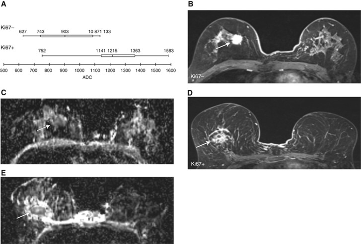
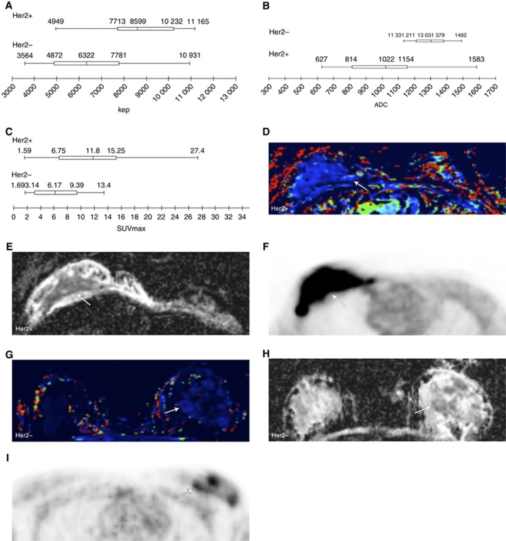
ER/PR- tumours demonstrated higher Kep and SUV than ER or PR+ tumours. HER2- tumours displayed higher ADC, Kep, and SUV than HER2+tumours. Only ADC discriminated Ki67⩽14% tumours (lower ADC) from Ki67>14% tumours. PET/MR biomarkers correlated with IHC phenotype in 13 out of 21 patients (62%; P=0.001).

CONCLUSIONS

Positron emission tomography magnetic resonance might non-invasively help discriminate IDC phenotypes, helping to optimise individual therapy options.

摘要

背景

浸润性导管癌(IDC)的基因和受体表达(表型)差异会影响预后和治疗反应。免疫组织化学(IHC)是IDC表型分析最常用的技术,但存在一些局限性,包括其侵入性。我们探讨了对比增强正电子发射断层扫描磁共振(CE-FDG PET/MR)鉴别IDC表型的可能性。

方法

21例接受了雌激素受体(ER)、孕激素受体(PR)、人表皮生长因子-2(HER2)和抗原Ki-67(Ki67)免疫组织化学评估的IDC患者接受了CE-FDG PET/MR检查。使用学生t检验和单因素方差分析,将磁共振灌注生物标志物、表观扩散系数(ADC)和标准摄取值(SUV)与免疫组织化学标志物及表型进行比较。

结果

ER/PR阴性肿瘤的 Kep 和 SUV 高于 ER 或 PR 阳性肿瘤。HER2 阴性肿瘤的 ADC、Kep 和 SUV 高于 HER2 阳性肿瘤。只有 ADC 能够区分 Ki67≤14%的肿瘤(较低的 ADC)和 Ki67>14%的肿瘤。21例患者中有13例(62%;P=0.001)的PET/MR生物标志物与免疫组织化学表型相关。

结论

正电子发射断层扫描磁共振可能有助于无创鉴别IDC表型,从而有助于优化个体化治疗方案。

https://cdn.ncbi.nlm.nih.gov/pmc/blobs/5cf5/5379139/20b3016d29c2/bjc201726f3.jpg
https://cdn.ncbi.nlm.nih.gov/pmc/blobs/5cf5/5379139/b6179ad5f1d6/bjc201726f1.jpg
https://cdn.ncbi.nlm.nih.gov/pmc/blobs/5cf5/5379139/4d6596dabe5a/bjc201726f2.jpg
https://cdn.ncbi.nlm.nih.gov/pmc/blobs/5cf5/5379139/20b3016d29c2/bjc201726f3.jpg
https://cdn.ncbi.nlm.nih.gov/pmc/blobs/5cf5/5379139/b6179ad5f1d6/bjc201726f1.jpg
https://cdn.ncbi.nlm.nih.gov/pmc/blobs/5cf5/5379139/4d6596dabe5a/bjc201726f2.jpg
https://cdn.ncbi.nlm.nih.gov/pmc/blobs/5cf5/5379139/20b3016d29c2/bjc201726f3.jpg

相似文献

1
PET/MR in invasive ductal breast cancer: correlation between imaging markers and histological phenotype.正电子发射断层显像/磁共振成像在浸润性导管癌中的应用:影像标志物与组织学表型的相关性
Br J Cancer. 2017 Mar 28;116(7):893-902. doi: 10.1038/bjc.2017.26. Epub 2017 Feb 16.
2
Associations between the standardized uptake value of (18)F-FDG PET/CT and the prognostic factors of invasive lobular carcinoma: in comparison with invasive ductal carcinoma.(18)F-FDG PET/CT标准化摄取值与浸润性小叶癌预后因素的相关性:与浸润性导管癌的比较
World J Surg Oncol. 2015 Mar 15;13:113. doi: 10.1186/s12957-015-0522-9.
3
Correlation of breast cancer subtypes, based on estrogen receptor, progesterone receptor, and HER2, with functional imaging parameters from ⁶⁸Ga-RGD PET/CT and ¹⁸F-FDG PET/CT.基于雌激素受体、孕激素受体和人表皮生长因子受体2的乳腺癌亚型与⁶⁸Ga-RGD PET/CT及¹⁸F-FDG PET/CT功能成像参数的相关性
Eur J Nucl Med Mol Imaging. 2014 Aug;41(8):1534-43. doi: 10.1007/s00259-014-2744-4. Epub 2014 Mar 21.
4
Diffusion-weighted imaging and FDG PET/CT: predicting the prognoses with apparent diffusion coefficient values and maximum standardized uptake values in patients with invasive ductal carcinoma.弥散加权成像和 FDG PET/CT:在浸润性导管癌患者中,通过表观扩散系数值和最大标准化摄取值预测预后。
World J Surg Oncol. 2012 Jun 28;10:126. doi: 10.1186/1477-7819-10-126.
5
Clinicopathological and prognostic relevance of uptake level using 18F-fluorodeoxyglucose positron emission tomography/computed tomography fusion imaging (18F-FDG PET/CT) in primary breast cancer.18F-氟脱氧葡萄糖正电子发射断层扫描/计算机断层扫描融合成像(18F-FDG PET/CT)摄取水平在原发性乳腺癌中的临床病理及预后相关性
Jpn J Clin Oncol. 2008 Apr;38(4):250-8. doi: 10.1093/jjco/hyn019.
6
A new method for apparent diffusion coefficient measurement using sequential (18)F-FDG PET and MRI: correlation with histological grade of invasive ductal carcinoma of the breast.一种使用连续(18)F-FDG PET 和 MRI 测量表观扩散系数的新方法:与乳腺浸润性导管癌组织学分级的相关性。
Ann Nucl Med. 2013 Oct;27(8):720-8. doi: 10.1007/s12149-013-0737-1. Epub 2013 May 23.
7
Relation between Ki-67, ER, PR, Her2/neu, p21, EGFR, and TOP II-α expression in invasive ductal breast cancer patients and correlations with prognosis.浸润性导管癌患者中Ki-67、雌激素受体(ER)、孕激素受体(PR)、人表皮生长因子受体2/neu(Her2/neu)、p21、表皮生长因子受体(EGFR)和拓扑异构酶II-α(TOP II-α)表达之间的关系及其与预后的相关性。
Asian Pac J Cancer Prev. 2015;16(2):823-9. doi: 10.7314/apjcp.2015.16.2.823.
8
Usefulness of 18F-FDG uptake with clinicopathologic and immunohistochemical prognostic factors in breast cancer.乳腺癌中 18F-FDG 摄取与临床病理及免疫组化预后因素的相关性。
Ann Nucl Med. 2012 Feb;26(2):175-83. doi: 10.1007/s12149-011-0556-1. Epub 2011 Dec 3.
9
Comparison of triple-negative and estrogen receptor-positive/progesterone receptor-positive/HER2-negative breast carcinoma using quantitative fluorine-18 fluorodeoxyglucose/positron emission tomography imaging parameters: a potentially useful method for disease characterization.使用定量氟-18氟脱氧葡萄糖/正电子发射断层扫描成像参数比较三阴性乳腺癌与雌激素受体阳性/孕激素受体阳性/人表皮生长因子受体2阴性乳腺癌:一种对疾病特征描述可能有用的方法。
Cancer. 2008 Mar 1;112(5):995-1000. doi: 10.1002/cncr.23226.
10
Prognostic significance of preoperative F-FDG PET/CT for breast cancer subtypes.术前F-FDG PET/CT对乳腺癌亚型的预后意义
Breast. 2016 Dec;30:5-12. doi: 10.1016/j.breast.2016.08.003. Epub 2016 Aug 29.

引用本文的文献

1
Microstructural diffusion MRI for differentiation of breast tumors and prediction of prognostic factors in breast cancer.用于鉴别乳腺肿瘤及预测乳腺癌预后因素的微观结构扩散磁共振成像
Front Oncol. 2025 Mar 5;15:1498691. doi: 10.3389/fonc.2025.1498691. eCollection 2025.
2
Diffusion-Weighted MRI for the Assessment of Molecular Prognostic Biomarkers in Breast Cancer.扩散加权磁共振成像在乳腺癌分子预后生物标志物评估中的应用。
Korean J Radiol. 2024 Jul;25(7):623-633. doi: 10.3348/kjr.2023.1188.
3
Joint EANM-SNMMI guideline on the role of 2-[F]FDG PET/CT in no special type breast cancer : (endorsed by the ACR, ESSO, ESTRO, EUSOBI/ESR, and EUSOMA).

本文引用的文献

1
Phase II trial of neoadjuvant letrozole and lapatinib in Asian postmenopausal women with estrogen receptor (ER) and human epidermal growth factor receptor 2 (HER2)-positive breast cancer [Neo-ALL-IN]: Highlighting the TILs, ER expressional change after neoadjuvant treatment, and FES-PET as potential significant biomarkers.亚洲绝经后雌激素受体(ER)和人表皮生长因子受体2(HER2)阳性乳腺癌患者新辅助来曲唑和拉帕替尼治疗的II期试验[Neo-ALL-IN]:强调肿瘤浸润淋巴细胞、新辅助治疗后ER表达变化以及FES-PET作为潜在的重要生物标志物
Cancer Chemother Pharmacol. 2016 Oct;78(4):685-95. doi: 10.1007/s00280-016-3107-6. Epub 2016 Aug 4.
2
Hybrid FDG-PET/MR compared to FDG-PET/CT in adult lymphoma patients.FDG-PET/MR 与 FDG-PET/CT 在成人淋巴瘤患者中的比较。
Abdom Radiol (NY). 2016 Jul;41(7):1338-48. doi: 10.1007/s00261-016-0638-6.
3
EANM-SNMMI 联合指南:2-[F]FDG PET/CT 在非特殊类型乳腺癌中的作用:(由 ACR、ESSO、ESTRO、EUSOBI/ESR 和 EUSOMA 认可)。
Eur J Nucl Med Mol Imaging. 2024 Jul;51(9):2706-2732. doi: 10.1007/s00259-024-06696-9. Epub 2024 May 14.
4
Prediction of therapy response of breast cancer patients with machine learning based on clinical data and imaging data derived from breast [F]FDG-PET/MRI.基于来自乳腺 [F]FDG-PET/MRI 的临床数据和影像学数据,应用机器学习预测乳腺癌患者的治疗反应。
Eur J Nucl Med Mol Imaging. 2024 Apr;51(5):1451-1461. doi: 10.1007/s00259-023-06513-9. Epub 2023 Dec 22.
5
Three-Dimensional In Vitro Tumor Spheroid Models for Evaluation of Anticancer Therapy: Recent Updates.用于评估抗癌治疗的三维体外肿瘤球体模型:最新进展
Cancers (Basel). 2023 Oct 4;15(19):4846. doi: 10.3390/cancers15194846.
6
Discordant and Converting Receptor Expressions in Brain Metastases from Breast Cancer: MRI-Based Non-Invasive Receptor Status Tracking.乳腺癌脑转移中不一致和转变的受体表达:基于MRI的非侵入性受体状态追踪
Cancers (Basel). 2023 May 23;15(11):2880. doi: 10.3390/cancers15112880.
7
Towards a fast PET/MRI protocol for breast cancer imaging: maintaining diagnostic confidence while reducing PET and MRI acquisition times.针对乳腺癌成像的快速 PET/MRI 协议:在减少 PET 和 MRI 采集时间的同时保持诊断信心。
Eur Radiol. 2023 Sep;33(9):6179-6188. doi: 10.1007/s00330-023-09580-6. Epub 2023 Apr 12.
8
Correlation between Imaging Markers Derived from PET/MRI and Invasive Acquired Biomarkers in Newly Diagnosed Breast Cancer.正电子发射断层扫描/磁共振成像衍生的影像标志物与新诊断乳腺癌侵袭性获得性生物标志物之间的相关性
Cancers (Basel). 2023 Mar 8;15(6):1651. doi: 10.3390/cancers15061651.
9
The influence of receptor expression and clinical subtypes on baseline [18F]FDG uptake in breast cancer: systematic review and meta-analysis.受体表达和临床亚型对乳腺癌基线[18F]FDG摄取的影响:系统评价和荟萃分析。
EJNMMI Res. 2023 Jan 23;13(1):5. doi: 10.1186/s13550-023-00953-y.
10
Simultaneous multislice diffusion-weighted imaging versus standard diffusion-weighted imaging in whole-body PET/MRI.全身 PET/MRI 中同时多层扩散加权成像与标准扩散加权成像的比较。
Eur Radiol. 2023 Apr;33(4):2536-2547. doi: 10.1007/s00330-022-09275-4. Epub 2022 Dec 3.
Melanoma, Version 2.2016, NCCN Clinical Practice Guidelines in Oncology.黑色素瘤临床实践指南(2016 年版),NCCN 肿瘤学临床实践指南。
J Natl Compr Canc Netw. 2016 Apr;14(4):450-73. doi: 10.6004/jnccn.2016.0051.
4
Pre-clinical evaluation of eight DOTA coupled gastrin-releasing peptide receptor (GRP-R) ligands for in vivo targeting of receptor-expressing tumors.八种DOTA偶联胃泌素释放肽受体(GRP-R)配体用于体内靶向表达该受体肿瘤的临床前评估。
EJNMMI Res. 2016 Dec;6(1):17. doi: 10.1186/s13550-016-0175-x. Epub 2016 Feb 20.
5
Perfusion Parameters in Dynamic Contrast-enhanced MRI and Apparent Diffusion Coefficient Value in Diffusion-weighted MRI:: Association with Prognostic Factors in Breast Cancer.动态对比增强磁共振成像中的灌注参数与扩散加权磁共振成像中的表观扩散系数值:与乳腺癌预后因素的关联
Acad Radiol. 2016 Apr;23(4):446-56. doi: 10.1016/j.acra.2015.12.011. Epub 2016 Feb 3.
6
Standardized Uptake Values from PET/MRI in Metastatic Breast Cancer: An Organ-based Comparison With PET/CT.正电子发射断层显像/磁共振成像(PET/MRI)在转移性乳腺癌中的标准化摄取值:与正电子发射断层显像/计算机断层扫描(PET/CT)的基于器官的比较
Breast J. 2016 May;22(3):264-73. doi: 10.1111/tbj.12569. Epub 2016 Feb 4.
7
Neoadjuvant Therapy for Early-Stage Breast Cancer: Current Practice, Controversies, and Future Directions.早期乳腺癌的新辅助治疗:当前实践、争议及未来方向
Oncology (Williston Park). 2015 Nov;29(11):828-38.
8
Diffusion-Weighted Imaging in 3.0 Tesla Breast MRI: Diagnostic Performance and Tumor Characterization Using Small Subregions vs. Whole Tumor Regions of Interest.3.0特斯拉乳腺磁共振成像中的扩散加权成像:使用小感兴趣子区域与整个肿瘤感兴趣区域的诊断性能及肿瘤特征分析
PLoS One. 2015 Oct 12;10(10):e0138702. doi: 10.1371/journal.pone.0138702. eCollection 2015.
9
Stratifying triple-negative breast cancer prognosis using 18F-FDG-PET/CT imaging.使用18F-FDG-PET/CT成像对三阴性乳腺癌预后进行分层。
Breast Cancer Res Treat. 2015 Oct;153(3):607-16. doi: 10.1007/s10549-015-3558-1. Epub 2015 Sep 7.
10
Clinical implications of the intrinsic molecular subtypes of breast cancer.乳腺癌内在分子亚型的临床意义。
Breast. 2015 Nov;24 Suppl 2:S26-35. doi: 10.1016/j.breast.2015.07.008. Epub 2015 Aug 5.