Department of Diagnostic and Interventional Radiology, University Dusseldorf, Medical Faculty, Moorenstrasse 5, D-40225, Dusseldorf, Germany.
High-Field and Hybrid MR Imaging, University Hospital Essen, University Duisburg-Essen, D-45147, Essen, Germany.
Eur Radiol. 2023 Sep;33(9):6179-6188. doi: 10.1007/s00330-023-09580-6. Epub 2023 Apr 12.
To investigate the diagnostic feasibility of a shortened breast PET/MRI protocol in breast cancer patients.
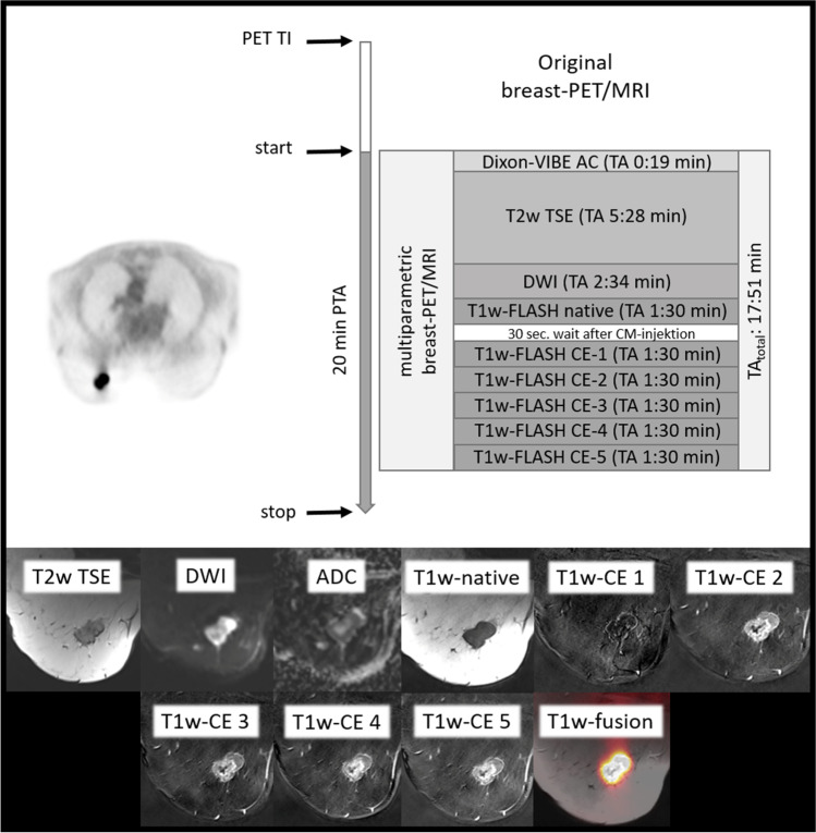
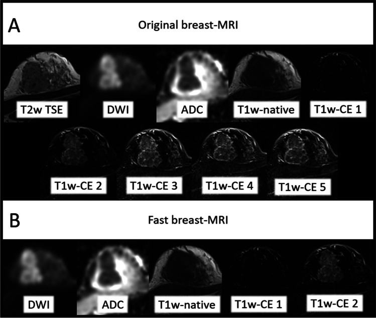
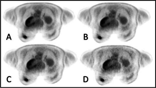
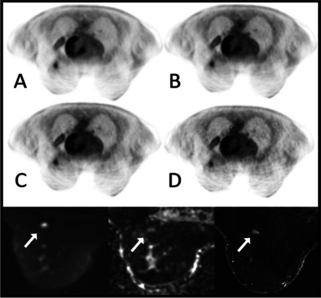
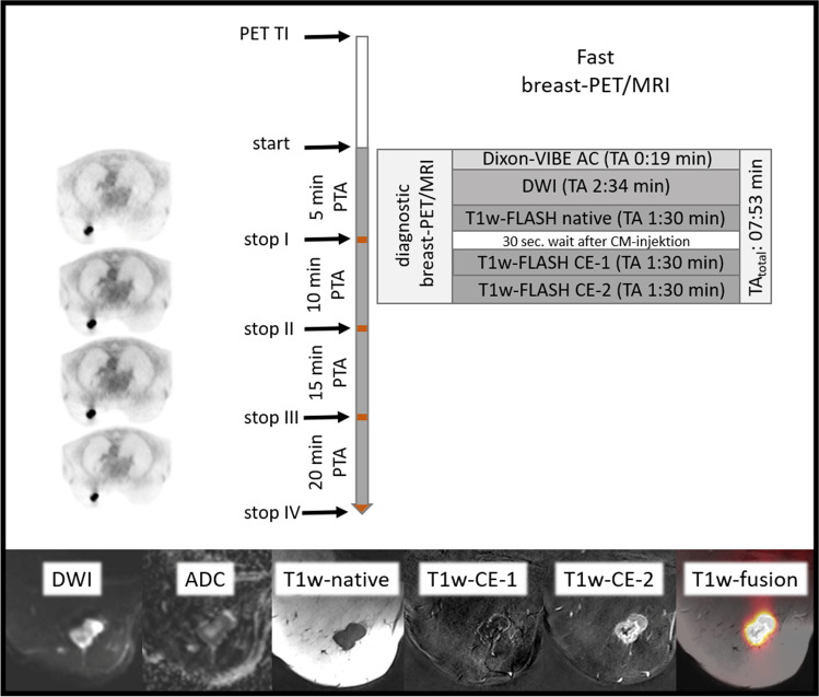
Altogether 90 women with newly diagnosed T1 (T1) and T2 (T2) breast cancer were included in this retrospective study. All underwent a dedicated comprehensive breast [F]FDG-PET/MRI. List-mode PET data were retrospectively reconstructed with 20, 15, 10, and 5 min for each patient to simulate the effect of reduced PET acquisition times. The SUV of all malign breast lesions was measured. Furthermore, breast PET data reconstructions were analyzed regarding image quality, lesion detectability, signal-to-noise ratio (SNR), and image noise (IN). The simultaneously acquired comprehensive MRI protocol was then shortened by retrospectively removing sequences from the protocol. Differences in malignant breast lesion detectability between the original and the fast breast MRI protocol were evaluated lesion-based. The 20-min PET reconstructions and the original MRI protocol served as reference.
In all PET reconstructions, 127 congruent breast lesions could be detected. Group comparison and T1 vs. T2 subgroup comparison revealed no significant difference of subjective image quality between 20, 15, 10, and 5 min acquisition times. SNR of qualitative image evaluation revealed no significant difference between different PET acquisition times. A slight but significant increase of IN with decreasing PET acquisition times could be detected. Lesion SUV group comparison between all PET acquisition times revealed no significant differences. Lesion-based evaluation revealed no significant difference in breast lesion detectability between original and fast breast MRI protocols.
Breast [F]FDG-PET/MRI protocols can be shortened from 20 to below 10 min without losing essential diagnostic information.
• A highly accurate breast cancer evaluation is possible by the shortened breast [F]FDG-PET/MRI examination protocol. • Significant time saving at breast [F]FDG-PET/MRI protocol could increase patient satisfaction and patient throughput for breast cancer patients at PET/MRI.
探讨缩短乳腺癌患者乳腺 PET/MRI 方案的诊断可行性。
本回顾性研究共纳入 90 例新诊断为 T1(T1)和 T2(T2)乳腺癌的女性患者。所有患者均接受了专门的综合乳腺 [F]FDG-PET/MRI 检查。为模拟减少 PET 采集时间的效果,对每位患者的列表模式 PET 数据进行回顾性重建,分别重建 20、15、10 和 5 分钟。测量所有恶性乳腺病变的 SUV。此外,还分析了乳腺 PET 数据重建的图像质量、病变检出率、信噪比(SNR)和图像噪声(IN)。然后通过从协议中回顾性地删除序列来缩短同时采集的综合 MRI 协议。基于病变评估了原始和快速乳腺 MRI 协议之间恶性乳腺病变检出率的差异。20 分钟 PET 重建和原始 MRI 协议作为参考。
在所有 PET 重建中,均能检测到 127 个一致的乳腺病变。组间比较和 T1 与 T2 亚组比较显示,不同 PET 采集时间的主观图像质量无显著差异。定性图像评估的 SNR 显示不同 PET 采集时间之间无显著差异。随着 PET 采集时间的减少,IN 略有但有统计学意义的增加。所有 PET 采集时间的病变 SUV 组间比较无显著差异。基于病变的评估显示,原始和快速乳腺 MRI 协议之间的乳腺病变检出率无显著差异。
乳腺 [F]FDG-PET/MRI 方案可从 20 分钟缩短至 10 分钟以下,而不会丢失重要的诊断信息。
• 缩短的乳腺 [F]FDG-PET/MRI 检查方案可实现对乳腺癌的高度准确评估。
• 在乳腺 [F]FDG-PET/MRI 方案中显著节省时间,可以提高乳腺癌患者的满意度和 PET/MRI 的患者吞吐量。