• 文献检索
  • 文档翻译
  • 深度研究
  • 学术资讯
  • Suppr Zotero 插件Zotero 插件
  • 邀请有礼
  • 套餐&价格
  • 历史记录
应用&插件
Suppr Zotero 插件Zotero 插件浏览器插件Mac 客户端Windows 客户端微信小程序
定价
高级版会员购买积分包购买API积分包
服务
文献检索文档翻译深度研究API 文档MCP 服务
关于我们
关于 Suppr公司介绍联系我们用户协议隐私条款
关注我们

Suppr 超能文献

核心技术专利:CN118964589B侵权必究
粤ICP备2023148730 号-1Suppr @ 2026

文献检索

告别复杂PubMed语法,用中文像聊天一样搜索,搜遍4000万医学文献。AI智能推荐,让科研检索更轻松。

立即免费搜索

文件翻译

保留排版,准确专业,支持PDF/Word/PPT等文件格式,支持 12+语言互译。

免费翻译文档

深度研究

AI帮你快速写综述,25分钟生成高质量综述,智能提取关键信息,辅助科研写作。

立即免费体验

花水母科及其他水螅水母亚纲动物(刺胞动物门,水螅纲)的外骨骼:结构与组成

Exoskeletons of Bougainvilliidae and other Hydroidolina (Cnidaria, Hydrozoa): structure and composition.

作者信息

Mendoza-Becerril María A, Marian José Eduardo A R, Migotto Alvaro Esteves, Marques Antonio Carlos

机构信息

Department of Zoology, Institute of Biosciences, University of São Paulo , São Paulo , Brazil.

Department of Zoology, Institute of Biosciences, University of São Paulo, São Paulo, Brazil; Center for Marine Biology, University of São Paulo, São Sebastião, São Paulo, Brazil.

出版信息

PeerJ. 2017 Feb 16;5:e2964. doi: 10.7717/peerj.2964. eCollection 2017.

DOI:10.7717/peerj.2964
PMID:28224050
原文链接:https://pmc.ncbi.nlm.nih.gov/articles/PMC5316283/
Abstract

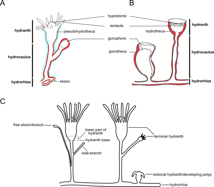
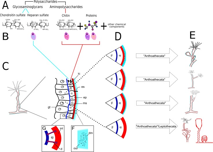
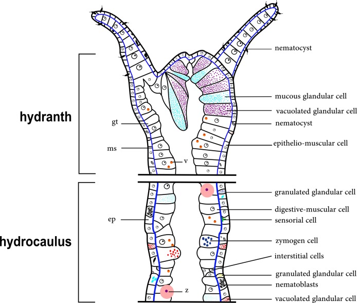
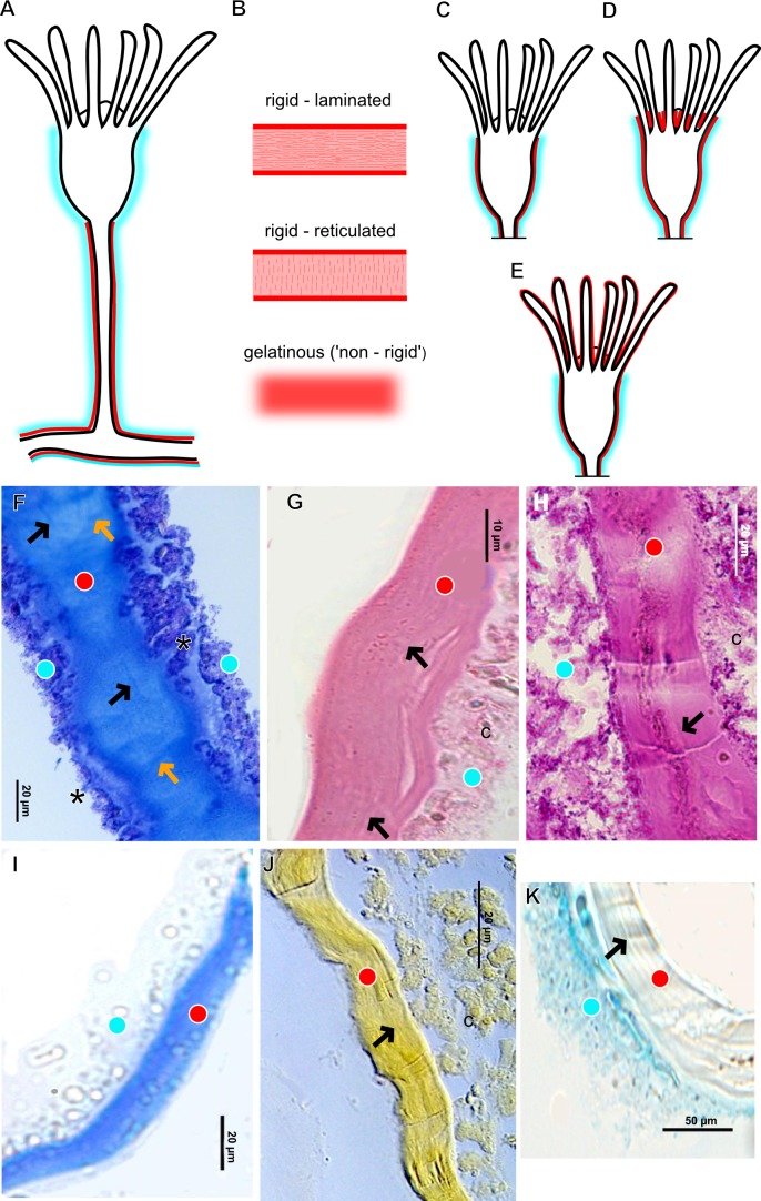
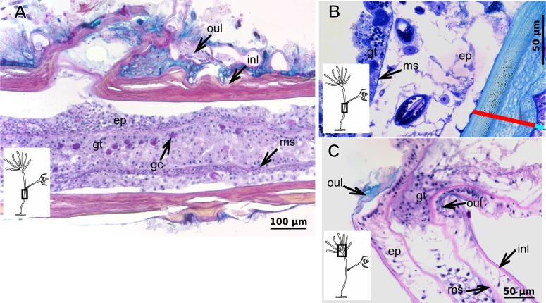
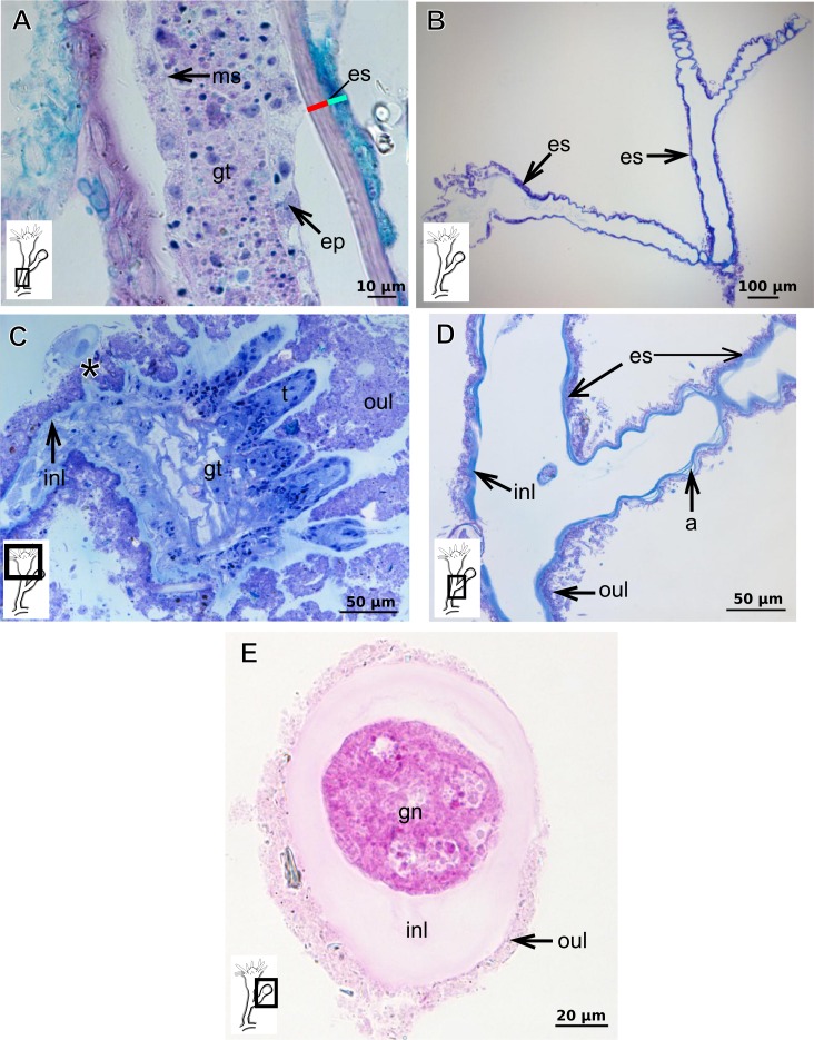
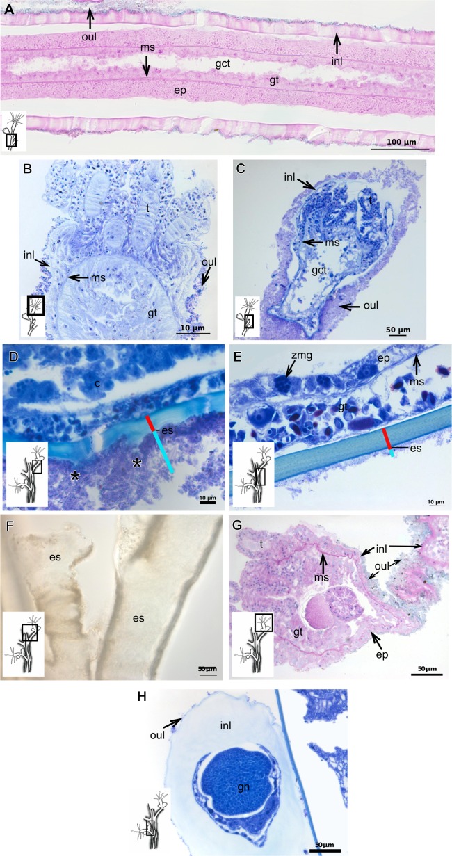
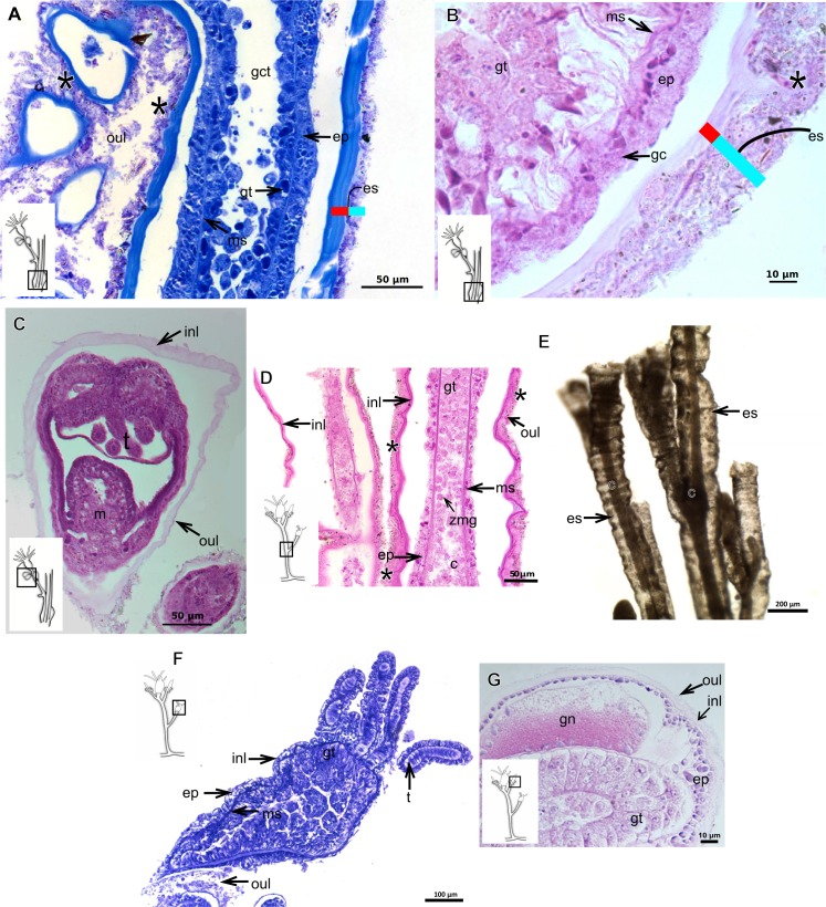
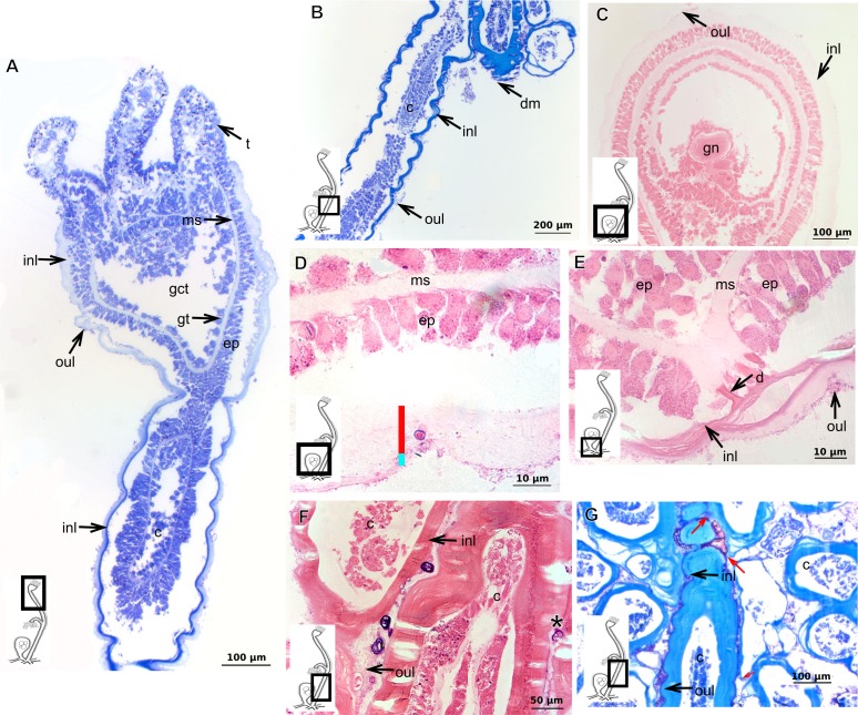
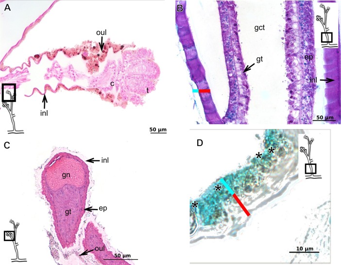
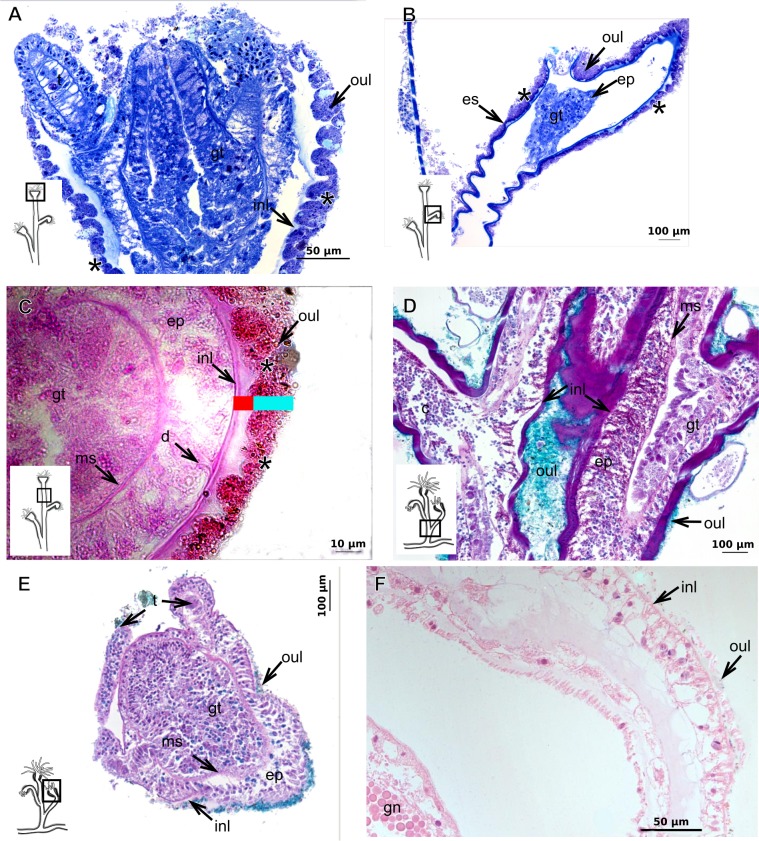
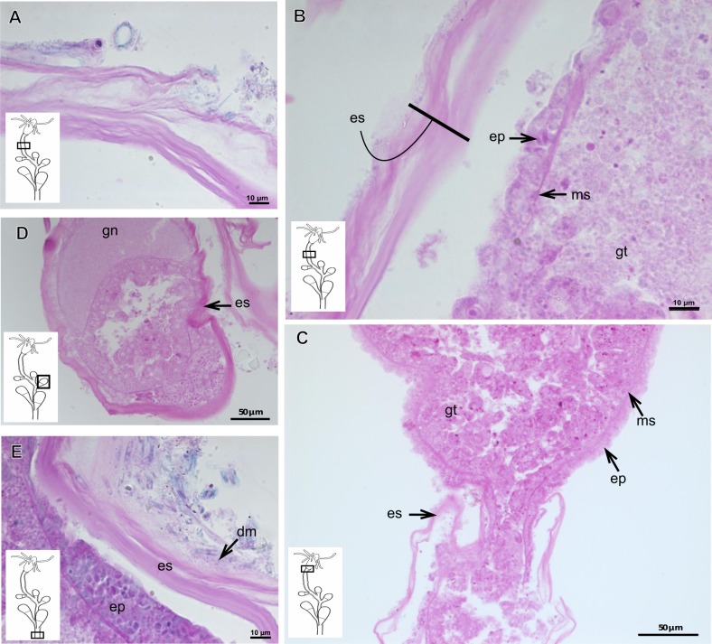
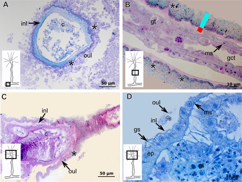
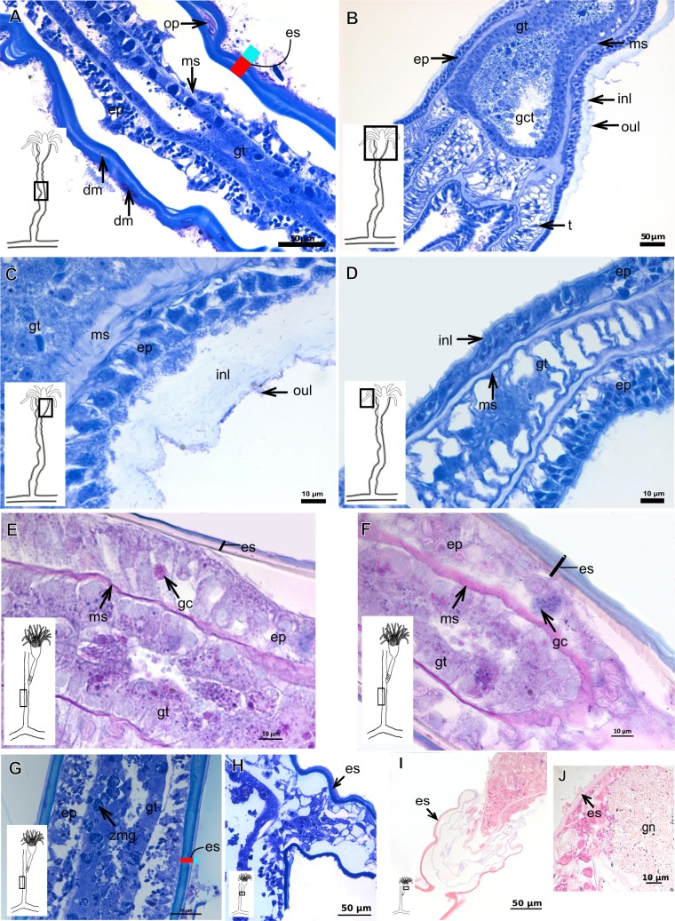
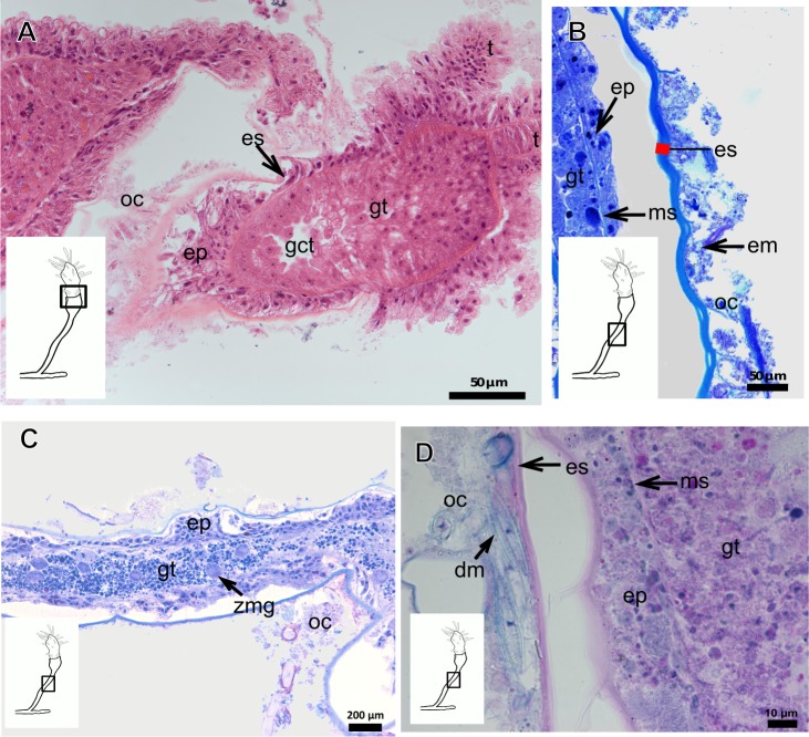
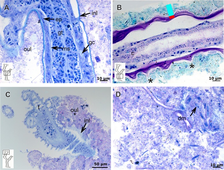
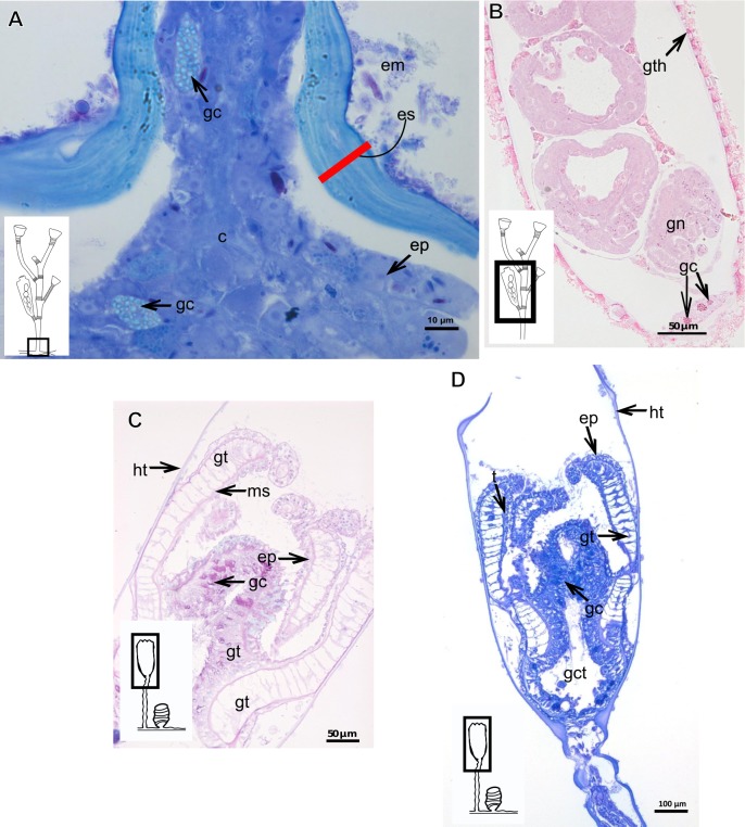
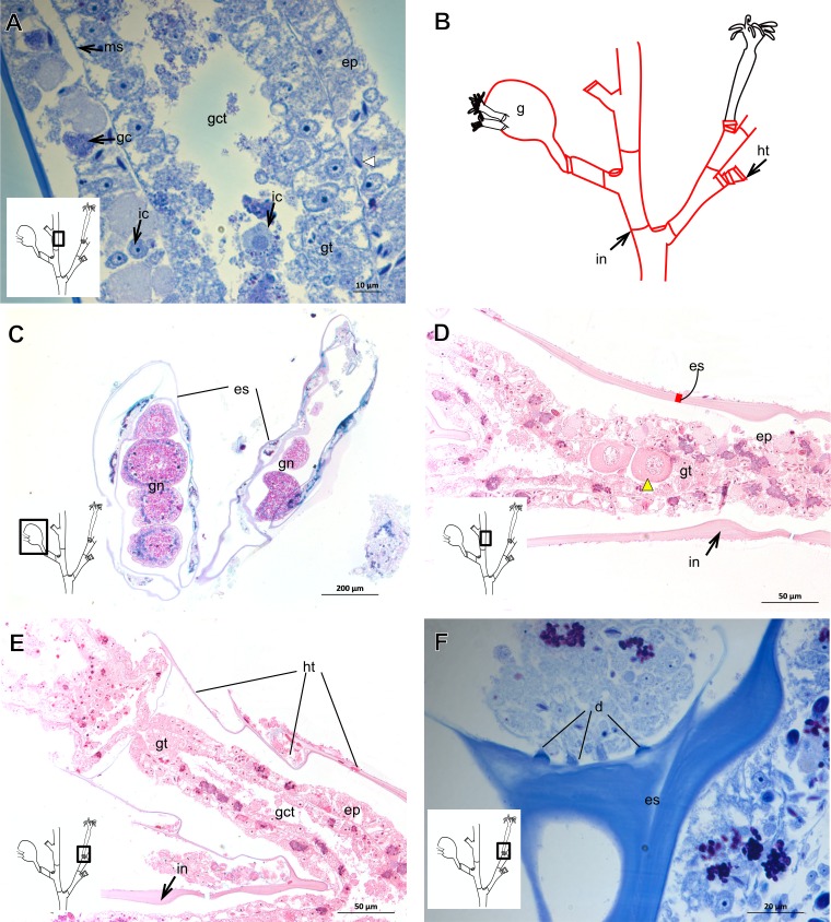
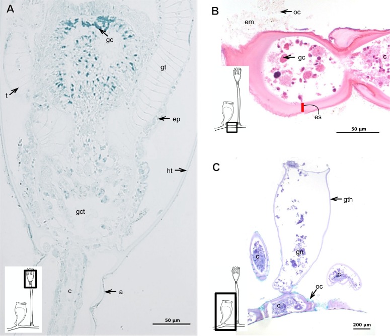
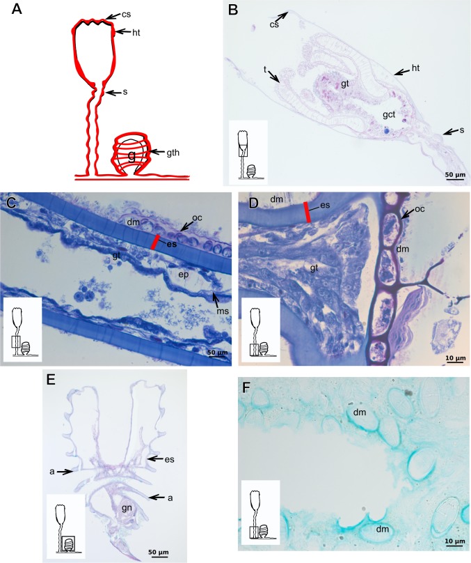
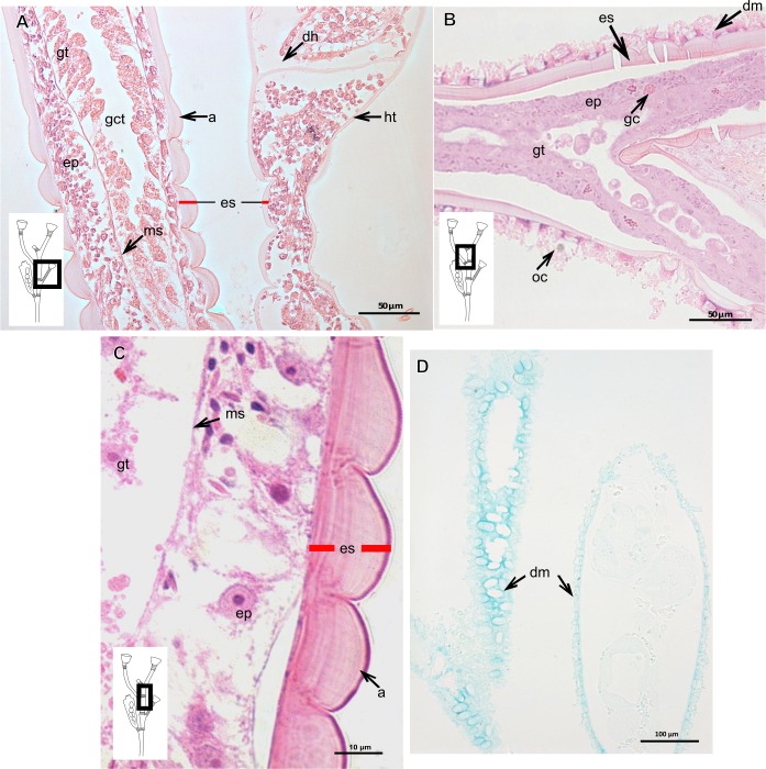
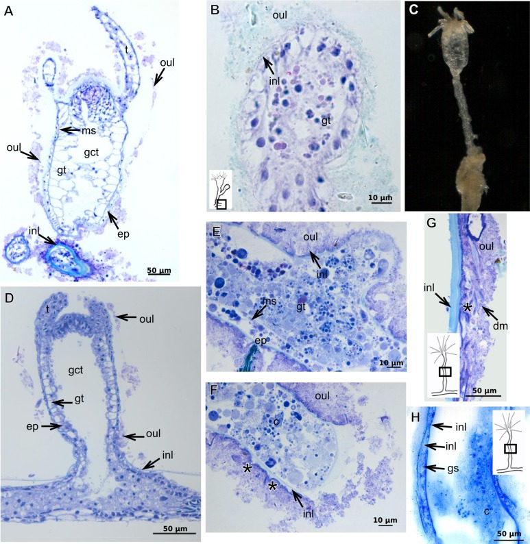
The exoskeleton is an important source of characters for the taxonomy of Hydroidolina. It originates as epidermal secretions and, among other functions, protects the coenosarc of the polypoid stage. However, comparative studies on the exoskeletal tissue origin, development, chemical, and structural characteristics, as well as its evolution and homology, are few and fragmented. This study compares the structure and composition of the exoskeleton and underlying coenosarc in members of "Anthoathecata" and some Leptothecata, but does so mainly in bougainvilliid polyps histological analyses. We also studied the development of the exoskeleton under experimental conditions. We identified three types of glandular epidermal cells related to the origin of the exoskeleton and the secretion of its polysaccharides component. The exoskeleton of the species studied is either bilayered (perisarc and exosarc, especially in bougainvilliids) or corneous (perisarc). The exoskeleton varies in chemical composition, structural rigidity, thickness, extension, and coverage in the different regions of the colony. In bilayered exoskeletons, the exosarc is produced first and appears to be a key step in the formation of the rigid exoskeleton. The exoskeleton contains anchoring structures such as desmocytes and "perisarc extensions."

摘要

外骨骼是水螅虫纲分类学中重要的特征来源。它起源于表皮分泌物,除其他功能外,还保护着螅形体阶段的共肉。然而,关于外骨骼组织的起源、发育、化学和结构特征,以及其进化和同源性的比较研究很少且零散。本研究比较了“花水母目”和一些软水母目的成员中外骨骼和其下方共肉的结构与组成,但主要是在布氏螅体的组织学分析中进行的。我们还在实验条件下研究了外骨骼的发育。我们确定了三种与外骨骼起源及其多糖成分分泌相关的腺状表皮细胞。所研究物种的外骨骼要么是双层的(围鞘和外鞘,特别是在布氏螅中),要么是角质的(围鞘)。外骨骼在群体的不同区域,其化学成分、结构硬度、厚度、延伸范围和覆盖程度各不相同。在双层外骨骼中,外鞘首先产生,似乎是形成坚硬外骨骼的关键步骤。外骨骼包含诸如连细胞和“围鞘延伸物”等锚定结构。

https://cdn.ncbi.nlm.nih.gov/pmc/blobs/d8e6/5316283/2e1e0e3c45b1/peerj-05-2964-g023.jpg
https://cdn.ncbi.nlm.nih.gov/pmc/blobs/d8e6/5316283/40be3cfa050d/peerj-05-2964-g001.jpg
https://cdn.ncbi.nlm.nih.gov/pmc/blobs/d8e6/5316283/ea85aab29939/peerj-05-2964-g002.jpg
https://cdn.ncbi.nlm.nih.gov/pmc/blobs/d8e6/5316283/5b3446287cd1/peerj-05-2964-g003.jpg
https://cdn.ncbi.nlm.nih.gov/pmc/blobs/d8e6/5316283/19bde88859f1/peerj-05-2964-g004.jpg
https://cdn.ncbi.nlm.nih.gov/pmc/blobs/d8e6/5316283/7d0775784e32/peerj-05-2964-g005.jpg
https://cdn.ncbi.nlm.nih.gov/pmc/blobs/d8e6/5316283/3f92b8552bd1/peerj-05-2964-g006.jpg
https://cdn.ncbi.nlm.nih.gov/pmc/blobs/d8e6/5316283/c46a1f1c3a8c/peerj-05-2964-g007.jpg
https://cdn.ncbi.nlm.nih.gov/pmc/blobs/d8e6/5316283/f4cf7a6551a2/peerj-05-2964-g008.jpg
https://cdn.ncbi.nlm.nih.gov/pmc/blobs/d8e6/5316283/5666f82d069c/peerj-05-2964-g009.jpg
https://cdn.ncbi.nlm.nih.gov/pmc/blobs/d8e6/5316283/e342abf0aa4e/peerj-05-2964-g010.jpg
https://cdn.ncbi.nlm.nih.gov/pmc/blobs/d8e6/5316283/7e160dda9cee/peerj-05-2964-g011.jpg
https://cdn.ncbi.nlm.nih.gov/pmc/blobs/d8e6/5316283/eee4be6258d2/peerj-05-2964-g012.jpg
https://cdn.ncbi.nlm.nih.gov/pmc/blobs/d8e6/5316283/7c0a78ff8751/peerj-05-2964-g013.jpg
https://cdn.ncbi.nlm.nih.gov/pmc/blobs/d8e6/5316283/b3a5b451f198/peerj-05-2964-g014.jpg
https://cdn.ncbi.nlm.nih.gov/pmc/blobs/d8e6/5316283/5e93ee76e67b/peerj-05-2964-g015.jpg
https://cdn.ncbi.nlm.nih.gov/pmc/blobs/d8e6/5316283/47bd22b30909/peerj-05-2964-g016.jpg
https://cdn.ncbi.nlm.nih.gov/pmc/blobs/d8e6/5316283/9b1fca2e7f25/peerj-05-2964-g017.jpg
https://cdn.ncbi.nlm.nih.gov/pmc/blobs/d8e6/5316283/6e67f5449a04/peerj-05-2964-g018.jpg
https://cdn.ncbi.nlm.nih.gov/pmc/blobs/d8e6/5316283/8d2d6c971be9/peerj-05-2964-g019.jpg
https://cdn.ncbi.nlm.nih.gov/pmc/blobs/d8e6/5316283/1f43401653fd/peerj-05-2964-g020.jpg
https://cdn.ncbi.nlm.nih.gov/pmc/blobs/d8e6/5316283/88ab66b1e18b/peerj-05-2964-g021.jpg
https://cdn.ncbi.nlm.nih.gov/pmc/blobs/d8e6/5316283/2e1e0e3c45b1/peerj-05-2964-g023.jpg

相似文献

1
Exoskeletons of Bougainvilliidae and other Hydroidolina (Cnidaria, Hydrozoa): structure and composition.花水母科及其他水螅水母亚纲动物(刺胞动物门,水螅纲)的外骨骼:结构与组成
PeerJ. 2017 Feb 16;5:e2964. doi: 10.7717/peerj.2964. eCollection 2017.
2
Anchoring cells (Desmocytes) in the hydrozoan polyp Cordylophora.淡水水螅体Cordylophora中的锚定细胞(间皮细胞)。
Tissue Cell. 1978;10(1):113-24. doi: 10.1016/0040-8166(78)90010-1.
3
Towards a phylogenetic classification of Leptothecata (Cnidaria, Hydrozoa).迈向软鞘水母亚纲(刺胞动物门,水螅纲)的系统发育分类
Sci Rep. 2016 Jan 29;6:18075. doi: 10.1038/srep18075.
4
Additions to the hydroids (Cnidaria, Hydrozoa) of the Bay of Fundy, northeastern North America, with a checklist of species reported from the region.北美东北部芬迪湾水螅虫纲(刺胞动物门,水螅虫目)物种新增记录及该地区已报道物种名录
Zootaxa. 2017 Apr 24;4256(1):1-86. doi: 10.11646/zootaxa.4256.1.1.
5
On a collection of hydroids (Cnidaria, Hydrozoa) from the southwest coast of Florida, USA.关于来自美国佛罗里达州西南海岸的水螅虫(刺胞动物门,水螅纲)的一个集合。
Zootaxa. 2019 Oct 25;4689(1):zootaxa.4689.1.1. doi: 10.11646/zootaxa.4689.1.1.
6
Some leptothecate hydroids (Cnidaria, Hydrozoa) from Hawaii, mostly from inshore and nearshore waters.一些来自夏威夷的具鞘水螅虫(刺胞动物门,水螅纲),大多来自近岸和近海海域。
Zootaxa. 2020 Aug 13;4830(2):zootaxa.4830.2.1. doi: 10.11646/zootaxa.4830.2.1.
7
Phylogenetic analysis of higher-level relationships within Hydroidolina (Cnidaria: Hydrozoa) using mitochondrial genome data and insight into their mitochondrial transcription.利用线粒体基因组数据对水螅水母纲(刺胞动物门:水螅虫纲)内高级别关系进行系统发育分析,并深入了解其线粒体转录情况。
PeerJ. 2015 Nov 19;3:e1403. doi: 10.7717/peerj.1403. eCollection 2015.
8
The genus L. Agassiz, 1862 (Cnidaria, Hydrozoa) in Northwest Africa.1862年的L. 阿加西属(刺胞动物门,水螅纲)在非洲西北部。
Zootaxa. 2017 Dec 12;4363(3):301-349. doi: 10.11646/zootaxa.4363.3.1.
9
Organizer regions in marine colonial hydrozoans.海洋群体水螅虫纲动物中的组织者区域。
Zoology (Jena). 2015 Apr;118(2):89-101. doi: 10.1016/j.zool.2014.12.001. Epub 2015 Feb 7.
10
New record of Hydrozoa Distichopora violacea (Pallas, 1766) (Anthoathecata: Stylasteridae) from Andaman and Nicobar Islands, India.印度安达曼和尼科巴群岛发现的紫枝多歧水母(Pallas,1766年)(花裸螅目:柱星螅科)新记录。
Zootaxa. 2018 Oct 31;4508(3):439-442. doi: 10.11646/zootaxa.4508.3.8.

引用本文的文献

1
A morpho-histological analysis of the exoskeleton of (Cnidaria, Hydrozoa) reveals insights into the taxonomy of Clathrozoellidae and Hydroidolina.对(刺胞动物门,水螅虫纲)外骨骼的形态组织学分析揭示了对笼螅科和水螅亚纲分类学的见解。
PeerJ. 2025 Jan 9;13:e18407. doi: 10.7717/peerj.18407. eCollection 2025.
2
A crown-group cnidarian from the Ediacaran of Charnwood Forest, UK.英国查恩伍德森林埃迪卡拉纪的一个冠群刺胞动物。
Nat Ecol Evol. 2022 Aug;6(8):1095-1104. doi: 10.1038/s41559-022-01807-x. Epub 2022 Jul 25.

本文引用的文献

1
Carbohydrates of the Organic Shell Matrix and the Shell-Forming Tissue of the Snail Biomphalaria glabrata (Say).光滑双脐螺(赛伊)有机壳基质和壳形成组织中的碳水化合物
Biol Bull. 1998 Apr;194(2):231-240. doi: 10.2307/1543052.
2
Phylogenetic relationships of Proboscoida Broch, 1910 (Cnidaria, Hydrozoa): Are traditional morphological diagnostic characters relevant for the delimitation of lineages at the species, genus, and family levels?1910年布罗奇的长吻水母目(刺胞动物门,水螅纲)的系统发育关系:传统形态学诊断特征对于物种、属和科级谱系的划分是否相关?
Mol Phylogenet Evol. 2017 Jan;106:118-135. doi: 10.1016/j.ympev.2016.09.012. Epub 2016 Sep 14.
3
Towards a phylogenetic classification of Leptothecata (Cnidaria, Hydrozoa).
迈向软鞘水母亚纲(刺胞动物门,水螅纲)的系统发育分类
Sci Rep. 2016 Jan 29;6:18075. doi: 10.1038/srep18075.
4
Reassessment of morphological diagnostic characters and species boundaries requires taxonomical changes for the genus orthopyxis L. Agassiz, 1862 (campanulariidae, hydrozoa) and some related campanulariids.对形态学诊断特征和物种界限的重新评估需要对1862年命名的Orthopyxis L. Agassiz属(桔梗科,水螅虫纲)以及一些相关桔梗科动物进行分类学修订。
PLoS One. 2015 Feb 27;10(2):e0117553. doi: 10.1371/journal.pone.0117553. eCollection 2015.
5
Growth and form in calyptoblastic hydroids; polymorphism within the Campanularidae.萼鞘水母的生长与形态;钟螅科内的多态性。
J Morphol. 1950 Jul;87(1):1-26. doi: 10.1002/jmor.1050870102.
6
Muscular anatomy of the Podocoryna carnea hydrorhiza.肉足纲有孔虫 Podocoryna carnea hydrorhiza 的肌肉解剖结构。
PLoS One. 2013 Aug 14;8(8):e72221. doi: 10.1371/journal.pone.0072221. eCollection 2013.
7
Marine hydroid perisarc: a chitin- and melanin-reinforced composite with DOPA-iron(III) complexes.海洋水螅体皮:一种富含几丁质和黑色素的复合物,含有 DOPA-铁(III)配合物。
Acta Biomater. 2013 Sep;9(9):8110-7. doi: 10.1016/j.actbio.2013.06.015. Epub 2013 Jun 19.
8
Horizontal gene transfer contributed to the evolution of extracellular surface structures: the freshwater polyp Hydra is covered by a complex fibrous cuticle containing glycosaminoglycans and proteins of the PPOD and SWT (sweet tooth) families.水平基因转移促进了细胞外表面结构的进化:淡水水螅被一层复杂的纤维状角质层覆盖,其中含有糖胺聚糖和 PPOD 和 SWT(甜牙)家族的蛋白质。
PLoS One. 2012;7(12):e52278. doi: 10.1371/journal.pone.0052278. Epub 2012 Dec 27.
9
Global diversity of the Stylasteridae (Cnidaria: Hydrozoa: Athecatae).全球石珊瑚科(刺胞动物门:水螅虫纲:无柄目)的多样性。
PLoS One. 2011;6(7):e21670. doi: 10.1371/journal.pone.0021670. Epub 2011 Jul 22.
10
Shared skeletal support in a coral-hydroid symbiosis.珊瑚-水螅共生体中的共享骨骼支持。
PLoS One. 2011;6(6):e20946. doi: 10.1371/journal.pone.0020946. Epub 2011 Jun 14.