Barrow Neurological Institute, 350 W Thomas Rd, Phoenix, AZ 85013, USA.
División de Neurociencias, Universidad Pablo de Olavide, Ctra. Utrera km. 1, 41013 Sevilla, Spain.
Sci Rep. 2017 Feb 27;7:43276. doi: 10.1038/srep43276.
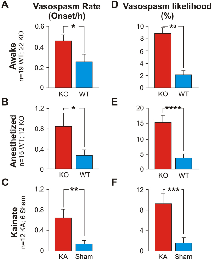
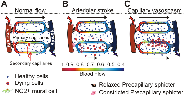
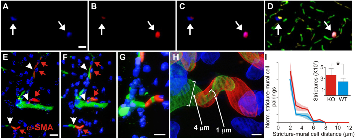
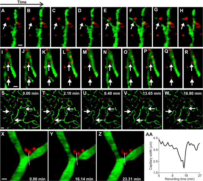
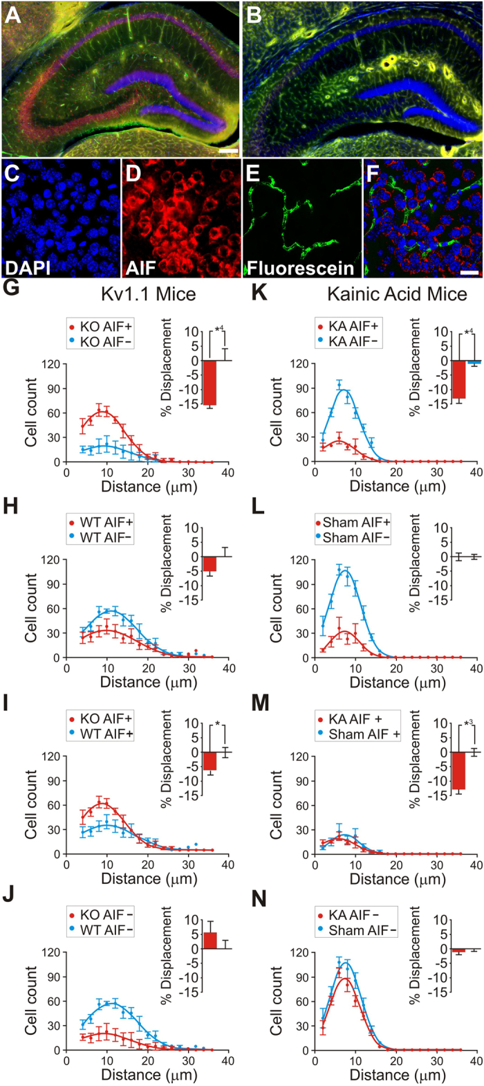
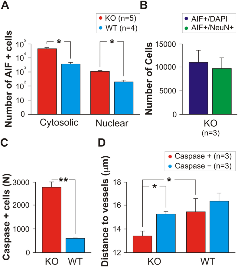
Seizure-driven brain damage in epilepsy accumulates over time, especially in the hippocampus, which can lead to sclerosis, cognitive decline, and death. Excitotoxicity is the prevalent model to explain ictal neurodegeneration. Current labeling technologies cannot distinguish between excitotoxicity and hypoxia, however, because they share common molecular mechanisms. This leaves open the possibility that undetected ischemic hypoxia, due to ictal blood flow restriction, could contribute to neurodegeneration previously ascribed to excitotoxicity. We tested this possibility with Confocal Laser Endomicroscopy (CLE) and novel stereological analyses in several models of epileptic mice. We found a higher number and magnitude of NG2+ mural-cell mediated capillary constrictions in the hippocampus of epileptic mice than in that of normal mice, in addition to spatial coupling between capillary constrictions and oxidative stressed neurons and neurodegeneration. These results reveal a role for hypoxia driven by capillary blood flow restriction in ictal neurodegeneration.
癫痫引起的脑损伤会随着时间的推移而积累,特别是在海马体中,这可能导致硬化、认知能力下降和死亡。兴奋性毒性是解释癫痫性神经退行性变的流行模型。然而,目前的标记技术无法区分兴奋性毒性和缺氧,因为它们具有共同的分子机制。这就使得由于癫痫发作时血流受限而未被检测到的缺血缺氧有可能导致以前归因于兴奋性毒性的神经退行性变。我们使用共聚焦激光内镜(CLE)和几种癫痫小鼠模型中的新的立体学分析对此进行了测试。我们发现,与正常小鼠相比,癫痫小鼠海马体中 NG2+壁细胞介导的毛细血管收缩的数量和幅度更高,此外,毛细血管收缩与氧化应激神经元和神经退行性变之间存在空间偶联。这些结果揭示了毛细血管血流受限引起的缺氧在癫痫性神经退行性变中的作用。