Kitahara Keisuke, Numako Chiya, Terada Yasuko, Nitta Kiyohumi, Shimada Yoshiya, Homma-Takeda Shino
Graduate School of Science, Chiba University, 1-33 Yayoi-cho, Inage-ku, Chiba 263-8522, Japan.
Japan Synchrotron Radiation Research Institute, Mikazuki, Hyogo 679-5198, Japan.
J Synchrotron Radiat. 2017 Mar 1;24(Pt 2):456-462. doi: 10.1107/S1600577517001850. Epub 2017 Feb 20.
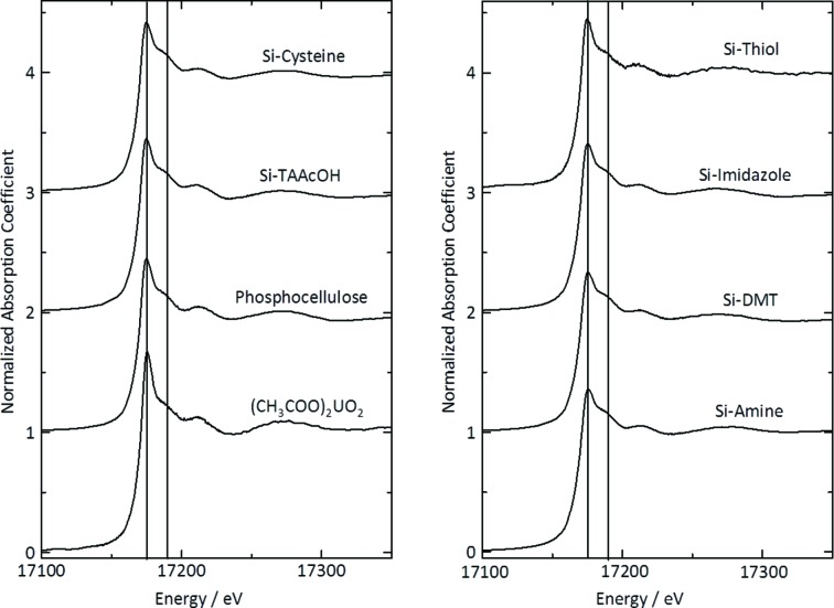
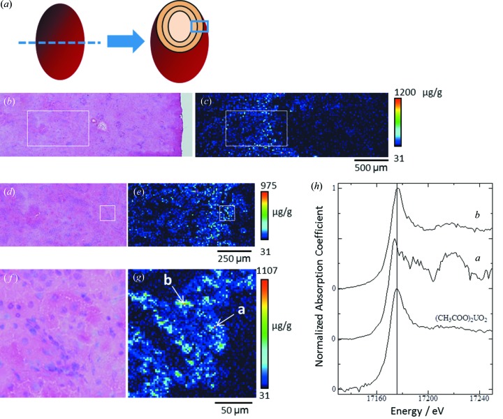
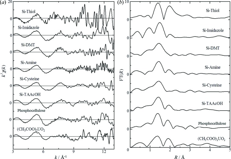
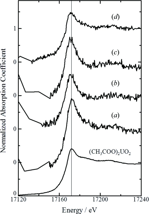
The kidney is the critical target of uranium exposure because uranium accumulates in the proximal tubules and causes tubular damage, but the chemical nature of uranium in kidney, such as its chemical status in the toxic target site, is poorly understood. Micro-X-ray absorption fine-structure (µXAFS) analysis was used to examine renal thin sections of rats exposed to uranyl acetate. The U L-edge X-ray absorption near-edge structure spectra of bulk renal specimens obtained at various toxicological phases were similar to that of uranyl acetate: their edge position did not shift compared with that of uranyl acetate (17.175 keV) although the peak widths for some kidney specimens were slightly narrowed. µXAFS measurements of spots of concentrated uranium in the micro-regions of the proximal tubules showed that the edge jump slightly shifted to lower energy. The results suggest that most uranium accumulated in kidney was uranium (VI) but a portion might have been biotransformed in rats exposed to uranyl acetate.
肾脏是铀暴露的关键靶器官,因为铀会在近端小管中蓄积并导致肾小管损伤,但是铀在肾脏中的化学性质,比如其在毒性靶位点的化学状态,目前还知之甚少。利用微X射线吸收精细结构(µXAFS)分析来检测暴露于醋酸双氧铀的大鼠的肾脏薄片。在不同毒理学阶段获取的大量肾脏标本的U L边X射线吸收近边结构光谱与醋酸双氧铀的光谱相似:尽管某些肾脏标本的峰宽略有变窄,但其边缘位置与醋酸双氧铀(17.175 keV)相比并未发生偏移。对近端小管微区中浓缩铀斑点的µXAFS测量表明,边缘跃变略微向低能量方向移动。结果表明,肾脏中蓄积的大部分铀是铀(VI),但在暴露于醋酸双氧铀的大鼠中,有一部分铀可能已经发生了生物转化。