Institut de Radioprotection et de Sûreté Nucléaire, PSE-SANTE/SESANE/LRSI, BP17, 92262, Fontenay-aux-Roses Cedex, France.
Anal Bioanal Chem. 2020 May;412(13):3113-3122. doi: 10.1007/s00216-020-02561-4. Epub 2020 Mar 20.
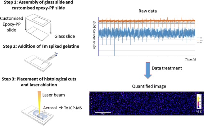
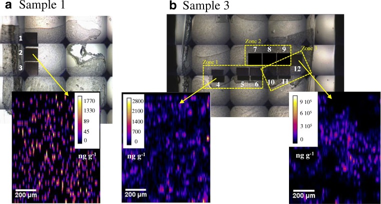
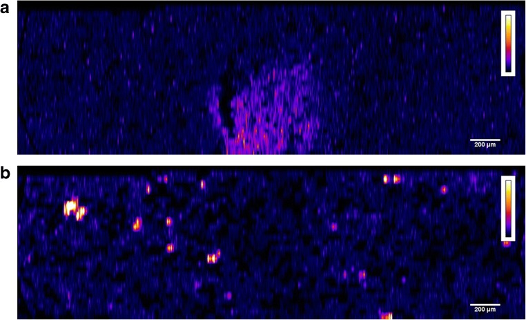
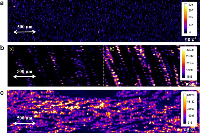
Mass spectrometry imaging (MSI) using laser ablation inductively coupled plasma mass spectrometry (LA-ICP-MS) has been employed for the elemental bio-distribution and quantification of uranium (U) in histological tissue sections of rodent kidneys. Kidneys were immediately immersed into 4% paraformaldehyde (PFA) solution for 24 h, Tissue-Tek O.C.T. Compound embedded and stored at - 80 °C until cutting in a cryostat, and mounted in gel-covered glass slides. In order to assure complete ablation of sample, sample preparation and laser conditions were carefully optimized. In this work, a new analytical methodology is presented for performing quantitative laser ablation analyses based on internal standard (thulium, Tm)-spiked gelatine (10% m/v) for correction of matrix effects, lack of tissue homogeneity, and instrumental drift. In parallel, matrix-matched laboratory standards, dosed at different concentrations of U, were prepared from a pool of rat kidneys. The quantitative images of cryo-sections revealed heterogeneous distribution of uranium within the renal tissue, because the cortical concentration was up to 120-fold higher than the medullary concentration. Graphical abstract.
利用激光烧蚀电感耦合等离子体质谱(LA-ICP-MS)的质谱成像(MSI)已被用于对啮齿动物肾脏组织切片中铀(U)的元素生物分布和定量。肾脏立即浸入 4%多聚甲醛(PFA)溶液中 24 小时,组织-Tek O.C.T. 化合物包埋并储存在-80°C 下直至在冷冻切片机中切割,并安装在凝胶覆盖的载玻片上。为了确保完全烧蚀样品,仔细优化了样品制备和激光条件。在这项工作中,提出了一种新的分析方法,用于基于内部标准(铥,Tm)掺杂明胶(10%m/v)进行定量激光烧蚀分析,以校正基质效应、组织不均匀性和仪器漂移。同时,从一组大鼠肾脏中制备了基质匹配的实验室标准品,剂量为不同浓度的 U。冷冻切片的定量图像显示铀在肾组织中的分布不均匀,因为皮质浓度比髓质浓度高 120 倍。