• 文献检索
  • 文档翻译
  • 深度研究
  • 学术资讯
  • Suppr Zotero 插件Zotero 插件
  • 邀请有礼
  • 套餐&价格
  • 历史记录
应用&插件
Suppr Zotero 插件Zotero 插件浏览器插件Mac 客户端Windows 客户端微信小程序
定价
高级版会员购买积分包购买API积分包
服务
文献检索文档翻译深度研究API 文档MCP 服务
关于我们
关于 Suppr公司介绍联系我们用户协议隐私条款
关注我们

Suppr 超能文献

核心技术专利:CN118964589B侵权必究
粤ICP备2023148730 号-1Suppr @ 2026

文献检索

告别复杂PubMed语法,用中文像聊天一样搜索,搜遍4000万医学文献。AI智能推荐,让科研检索更轻松。

立即免费搜索

文件翻译

保留排版,准确专业,支持PDF/Word/PPT等文件格式,支持 12+语言互译。

免费翻译文档

深度研究

AI帮你快速写综述,25分钟生成高质量综述,智能提取关键信息,辅助科研写作。

立即免费体验

通过锥形束计算机断层扫描(CBCT)图像评估颞下颌关节解剖结构。

Temporomandibular Joint Anatomy Assessed by CBCT Images.

作者信息

Caruso Silvia, Storti Ennio, Nota Alessandro, Ehsani Shideh, Gatto Roberto

机构信息

Department of Life, Health & Environmental Sciences, University of L'Aquila, L'Aquila, Italy.

Dental School, Vita-Salute San Raffaele University, Milan, Italy.

出版信息

Biomed Res Int. 2017;2017:2916953. doi: 10.1155/2017/2916953. Epub 2017 Feb 2.

DOI:10.1155/2017/2916953
PMID:28261607
原文链接:https://pmc.ncbi.nlm.nih.gov/articles/PMC5312052/
Abstract

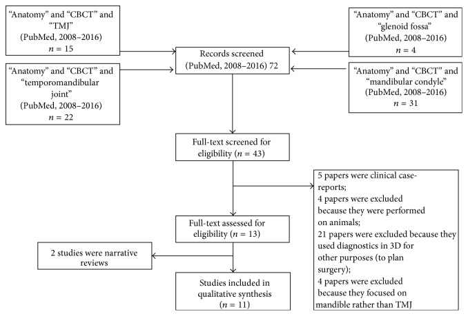
. Since cone beam computed tomography (CBCT) has been used for the study of craniofacial morphology, the attention of orthodontists has also focused on the mandibular condyle. The purpose of this brief review is to summarize the recent 3D CBCT images of mandibular condyle. . The eligibility criteria for the studies are (a) studies aimed at evaluating the anatomy of the temporomandibular joint; (b) studies performed with CBCT images; (c) studies on human subjects; (d) studies that were not clinical case-reports and clinical series; (e) studies reporting data on children, adolescents, or young adults (data from individuals with age ≤ 30 years). Sources included PubMed from June 2008 to June 2016. . 43 full-text articles were initially screened for eligibility. 13 full-text articles were assessed for eligibility. 11 articles were finally included in qualitative synthesis. The main topics treated in the studies are the volume and surface of the mandibular condyle, the bone changes on cortical surface, the facial asymmetry, and the optimum position of the condyle in the glenoid fossa. . Additional studies will be necessary in the future, constructed with longitudinal methodology, especially in growing subjects. The limits of CBCT acquisitions are also highlighted.

摘要

自从锥形束计算机断层扫描(CBCT)被用于颅面形态学研究以来,正畸医生的注意力也集中在了下颌髁突上。这篇简短综述的目的是总结下颌髁突最近的三维CBCT图像。. 纳入研究的标准为:(a)旨在评估颞下颌关节解剖结构的研究;(b)使用CBCT图像进行的研究;(c)针对人类受试者的研究;(d)非临床病例报告和临床系列研究;(e)报告儿童、青少年或年轻人(年龄≤30岁个体的数据)数据的研究。资料来源包括2008年6月至2016年6月的PubMed。. 最初筛选了43篇全文文章以确定其是否符合纳入标准。评估了13篇全文文章是否符合纳入标准。最终11篇文章被纳入定性综合分析。研究中涉及的主要主题是下颌髁突的体积和表面、皮质表面的骨质变化、面部不对称以及髁突在关节窝中的最佳位置。. 未来有必要采用纵向研究方法开展更多研究,尤其是针对生长发育期受试者的研究。同时也强调了CBCT采集的局限性。

https://cdn.ncbi.nlm.nih.gov/pmc/blobs/3eeb/5312052/5b348cb34f07/BMRI2017-2916953.001.jpg

相似文献

1
Temporomandibular Joint Anatomy Assessed by CBCT Images.通过锥形束计算机断层扫描(CBCT)图像评估颞下颌关节解剖结构。
Biomed Res Int. 2017;2017:2916953. doi: 10.1155/2017/2916953. Epub 2017 Feb 2.
2
Improved Visualization and Assessment of Condylar Position in the Glenoid Fossa for Different Occlusions: A CBCT Study.不同咬合情况下关节窝内髁突位置的可视化及评估改善:一项锥形束CT研究
J Contemp Dent Pract. 2016 Aug 1;17(8):679-86. doi: 10.5005/jp-journals-10024-1912.
3
Evaluation of temporomandibular fossa and mandibular condyle in adolescent patients affected by bilateral cleft lip and palate using cone beam computed tomography.使用锥形束计算机断层扫描评估双侧唇腭裂青少年患者的颞下颌窝和下颌髁突。
Scanning. 2016 Nov;38(6):720-726. doi: 10.1002/sca.21320. Epub 2016 Apr 22.
4
Effects of condylar head surface changes on mandibular position in patients with temporomandibular joint osteoarthritis.髁突头部表面变化对颞下颌关节骨关节炎患者下颌位置的影响。
J Craniomaxillofac Surg. 2015 Oct;43(8):1380-3. doi: 10.1016/j.jcms.2015.06.031. Epub 2015 Jun 27.
5
Bony deviations revealed by cone beam computed tomography of the temporomandibular joint in subjects without ongoing pain.无持续性疼痛患者的颞下颌关节锥形束 CT 检查所显示的骨性偏斜。
J Oral Facial Pain Headache. 2014 Fall;28(4):331-7. doi: 10.11607/ofph.1255.
6
Short- and long-term changes of condylar position after bilateral sagittal split ramus osteotomy for mandibular advancement in combination with Le Fort I osteotomy evaluated by cone-beam computed tomography.通过锥形束计算机断层扫描评估下颌前徙联合Le Fort I截骨术的双侧矢状劈开下颌支截骨术后髁突位置的短期和长期变化。
J Oral Maxillofac Surg. 2013 Nov;71(11):1956-66. doi: 10.1016/j.joms.2013.06.213. Epub 2013 Aug 22.
7
Association between mandibular condylar position and clinical dysfunction index.下颌髁突位置与临床功能障碍指数之间的关联。
J Craniomaxillofac Surg. 2015 May;43(4):432-6. doi: 10.1016/j.jcms.2015.01.005. Epub 2015 Jan 24.
8
Three-dimensional analysis of the temporomandibular joint and fossa-condyle relationship.颞下颌关节及关节窝-髁突关系的三维分析
Orthodontics (Chic.). 2011 Fall;12(3):210-21.
9
Radiographic examination of the temporomandibular joint using cone beam computed tomography.使用锥形束计算机断层扫描对颞下颌关节进行影像学检查。
Dentomaxillofac Radiol. 2004 May;33(3):196-201. doi: 10.1259/dmfr/27403192.
10
[Change in condylar and mandibular morphology in juvenile idiopathic arthritis: cone beam volumetric imaging].青少年特发性关节炎中髁突及下颌骨形态的改变:锥形束容积成像
Fogorv Sz. 2013 Mar;106(1):27-31.

引用本文的文献

1
Modifications of the articular eminence inclination in association with the loss of occlusal support in the molar area. A cone-beam computed tomography study.髁突前斜面倾斜度的改变与磨牙后区咬合支持丧失的相关性:锥形束 CT 研究。
Rom J Morphol Embryol. 2024 Apr-Jun;65(2):297-307. doi: 10.47162/RJME.65.2.17.
2
Appraisal of the Accuracy and Reliability of Cone-Beam Computed Tomography and Three-Dimensional Printing for Volumetric Mandibular Condyle Measurements of a Human Condyle.锥形束计算机断层扫描和三维打印用于人体髁突下颌骨髁突体积测量的准确性和可靠性评估。
Cureus. 2023 Oct 9;15(10):e46746. doi: 10.7759/cureus.46746. eCollection 2023 Oct.
3

本文引用的文献

1
Morphologic Analysis of the Temporomandibular Joint Between Patients With Facial Asymmetry and Asymptomatic Subjects by 2D and 3D Evaluation: A Preliminary Study.通过二维和三维评估对面部不对称患者与无症状受试者的颞下颌关节进行形态学分析:一项初步研究。
Medicine (Baltimore). 2016 Mar;95(13):e3052. doi: 10.1097/MD.0000000000003052.
2
Evaluation of mandibular condyles in children with unilateral posterior crossbite.评价单侧后牙反颌儿童的下颌髁突。
Braz Oral Res. 2015;29:49. doi: 10.1590/1807-3107BOR-2015.vol29.0049. Epub 2015 Mar 10.
3
Detection accuracy of condylar defects in cone beam CT images scanned with different resolutions and units.
Occlusal Splint Therapy Combined with Cranio-Temporomandibular Kinesiotherapy in Patients with Temporomandibular Disorders-A CBCT Study.
咬合板治疗联合颅颞下颌运动疗法治疗颞下颌关节紊乱病患者的CBCT研究
Life (Basel). 2022 Dec 19;12(12):2143. doi: 10.3390/life12122143.
4
Association between Psychological Status and Condylar Bony Changes in Adults: A Retrospective Survey-Based Study.成人心理状态与髁突骨质改变之间的关联:一项基于回顾性调查的研究
J Clin Med. 2022 Dec 17;11(24):7497. doi: 10.3390/jcm11247497.
5
Evaluation of the Mandibular Condyle Morphologic Relation before and after Orthognathic Surgery in Class II and III Malocclusion Patients Using Cone Beam Computed Tomography.使用锥形束计算机断层扫描评估安氏II类和III类错牙合患者正颌手术前后下颌髁突的形态学关系。
Biology (Basel). 2022 Sep 14;11(9):1353. doi: 10.3390/biology11091353.
6
Cone-Beam Computed Tomographic Assessment of the Mandibular Condylar Volume in Different Skeletal Patterns: A Retrospective Study in Adult Patients.不同骨骼类型下颌髁突体积的锥形束计算机断层扫描评估:一项针对成年患者的回顾性研究
Bioengineering (Basel). 2022 Mar 2;9(3):102. doi: 10.3390/bioengineering9030102.
7
Automated cortical thickness measurement of the mandibular condyle head on CBCT images using a deep learning method.使用深度学习方法在锥形束计算机断层扫描(CBCT)图像上自动测量下颌髁突头部的皮质厚度。
Sci Rep. 2021 Jul 21;11(1):14852. doi: 10.1038/s41598-021-94362-7.
8
Effects of Exposure Parameters and Voxel Size for Cone-Beam Computed Tomography on the Image Matching Accuracy with an Optical Dental Scan Image: An In Vitro Study.锥形束计算机断层扫描的曝光参数和体素大小对与光学牙科扫描图像的图像匹配精度的影响:一项体外研究。
Biomed Res Int. 2021 Jun 10;2021:6971828. doi: 10.1155/2021/6971828. eCollection 2021.
9
Three-Dimensional Assessment of Mandibular Condylar Volume and Position Subsequent to Twin Block Functional Therapy of Skeletal Class II Malocclusion Accompanied by Low-Level Laser Therapy.伴有低强度激光治疗的安氏II类错牙合畸形双期阻断性功能性矫治后髁突体积与位置的三维评估
Dent J (Basel). 2020 Oct 9;8(4):115. doi: 10.3390/dj8040115.
10
Morphology of the Temporomandibular Joints Regarding the Presence of Osteoarthritic Changes.颞下颌关节形态与骨关节炎变化的关系。
Int J Environ Res Public Health. 2020 Apr 23;17(8):2923. doi: 10.3390/ijerph17082923.
不同分辨率和单位的锥形束 CT 图像中髁突缺损的检测准确性。
Dentomaxillofac Radiol. 2014;43(3):20130414. doi: 10.1259/dmfr.20130414. Epub 2014 Jan 22.
4
TMJ imaging by CBCT: Current scenario.锥形束计算机断层扫描(CBCT)对颞下颌关节的成像:当前现状
Ann Maxillofac Surg. 2013 Jan;3(1):80-3. doi: 10.4103/2231-0746.110069.
5
Condylar volume and condylar area in class I, class II and class III young adult subjects.青少年 I 类、II 类和 III 类人群髁突体积和面积的研究。
Head Face Med. 2012 Dec 14;8:34. doi: 10.1186/1746-160X-8-34.
6
Do skeletal cephalometric characteristics correlate with condylar volume, surface and shape? A 3D analysis.颅面骨骼的形态特征是否与髁突体积、表面和形态相关?一项三维分析。
Head Face Med. 2012 May 15;8:15. doi: 10.1186/1746-160X-8-15.
7
Evaluation of bone changes in the temporomandibular joint using cone beam CT.使用锥形束 CT 评估颞下颌关节的骨变化。
Dentomaxillofac Radiol. 2012 Jan;41(1):24-9. doi: 10.1259/dmfr/17815139.
8
Assessment of optimal condylar position in the coronal and axial planes with limited cone-beam computed tomography.利用有限锥形束计算机断层扫描评估冠状面和轴位面的最佳髁突位置。
J Prosthodont. 2011 Aug;20(6):432-8. doi: 10.1111/j.1532-849X.2011.00730.x. Epub 2011 Jun 20.
9
Condylar volume and surface in Caucasian young adult subjects.高加索年轻成年人髁突体积和表面。
BMC Med Imaging. 2010 Dec 31;10:28. doi: 10.1186/1471-2342-10-28.
10
Assessment of simulated mandibular condyle bone lesions by cone beam computed tomography.锥形束计算机断层扫描评估模拟下颌髁突骨病变。
Braz Oral Res. 2010 Oct-Dec;24(4):467-74. doi: 10.1590/s1806-83242010000400016.