Barnoy Shoshana, Gancz Hanan, Zhu Yuewei, Honnold Cary L, Zurawski Daniel V, Venkatesan Malabi M
a Department of Enteric Infections , Bacterial Diseases Branch (BDB), Walter Reed Army Institute of Research , Silver Spring , Maryland , USA.
b Wound Infections Department , BDB, Walter Reed Army Institute of Research , Silver Spring Maryland , USA.
Gut Microbes. 2017 Jul 4;8(4):335-350. doi: 10.1080/19490976.2017.1293225. Epub 2017 Feb 13.
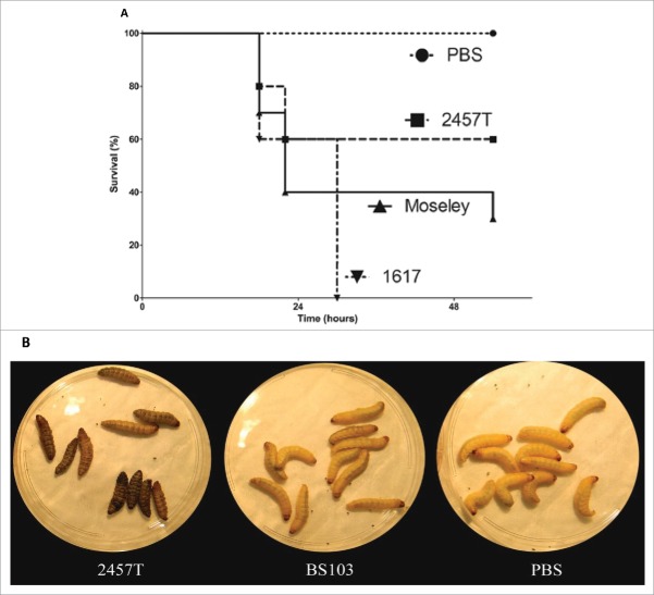
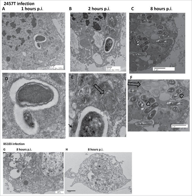
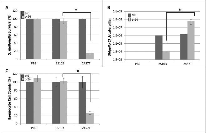
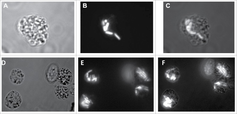
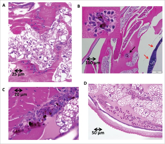
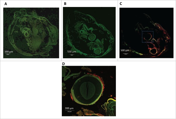
Shigella spp. causing bacterial diarrhea and dysentery are human enteroinvasive bacterial pathogens that are orally transmitted through contaminated food and water and cause bacillary dysentery. Although natural Shigella infections are restricted to humans and primates, several smaller animal models are used to analyze individual steps in pathogenesis. No animal model fully duplicates the human response and sustaining the models requires expensive animals, costly maintenance of animal facilities, veterinary services and approved animal protocols. This study proposes the development of the caterpillar larvae of Galleria mellonella as a simple, inexpensive, informative, and rapid in-vivo model for evaluating virulence and the interaction of Shigella with cells of the insect innate immunity. Virulent Shigella injected through the forelegs causes larvae death. The mortality rates were dependent on the Shigella strain, the infectious dose, and the presence of the virulence plasmid. Wild-type S. flexneri 2a, persisted and replicated within the larvae, resulting in haemocyte cell death, whereas plasmid-cured mutants were rapidly cleared. Histology of the infected larvae in conjunction with fluorescence, immunofluorescence, and transmission electron microscopy indicate that S. flexneri reside within a vacuole of the insect haemocytes that ultrastructurally resembles vacuoles described in studies with mouse and human macrophage cell lines. Some of these bacteria-laden vacuoles had double-membranes characteristic of autophagosomes. These results suggest that G. mellonella larvae can be used as an easy-to-use animal model to understand Shigella pathogenesis that requires none of the time and labor-consuming procedures typical of other systems.
引起细菌性腹泻和痢疾的志贺氏菌属是人类肠道侵袭性细菌病原体,通过受污染的食物和水经口传播,可导致杆菌性痢疾。虽然自然感染志贺氏菌的情况仅限于人类和灵长类动物,但一些小型动物模型被用于分析发病机制的各个步骤。没有一种动物模型能完全复制人类的反应,维持这些模型需要昂贵的动物、高昂的动物设施维护成本、兽医服务以及经过批准的动物实验方案。本研究提出将大蜡螟幼虫开发为一种简单、廉价、信息丰富且快速的体内模型,用于评估志贺氏菌的毒力以及志贺氏菌与昆虫先天免疫细胞的相互作用。通过前腿注射的有毒志贺氏菌会导致幼虫死亡。死亡率取决于志贺氏菌菌株、感染剂量以及毒力质粒的存在情况。野生型福氏志贺氏菌2a在幼虫体内持续存在并繁殖,导致血细胞死亡,而质粒缺失突变体则被迅速清除。对感染幼虫进行组织学检查,并结合荧光、免疫荧光和透射电子显微镜观察表明,福氏志贺氏菌存在于昆虫血细胞的液泡中,其超微结构类似于在小鼠和人类巨噬细胞系研究中描述的液泡。其中一些含有细菌的液泡具有自噬体的双膜特征。这些结果表明,大蜡螟幼虫可作为一种易于使用的动物模型,用于理解志贺氏菌的发病机制,而无需其他系统所特有的耗时费力的程序。