CRUK and EPSRC Imaging Centre, Division of Radiotherapy and Imaging, Institute of Cancer Research, Sutton, Surrey, UK.
Joint Department of Physics, Institute of Cancer Research and Royal Marsden NHS Foundation Trust, Sutton, Surrey, UK.
Sci Rep. 2017 Mar 13;7(1):165. doi: 10.1038/s41598-017-00144-5.
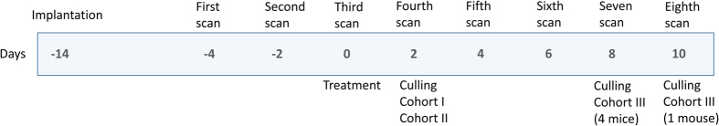
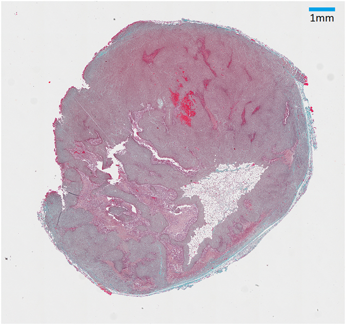
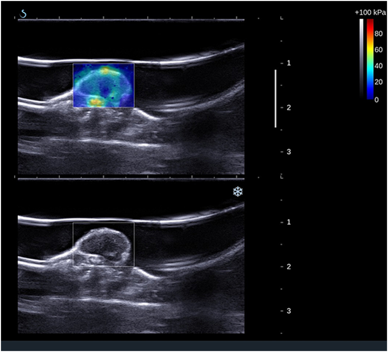
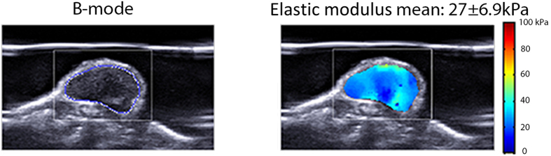
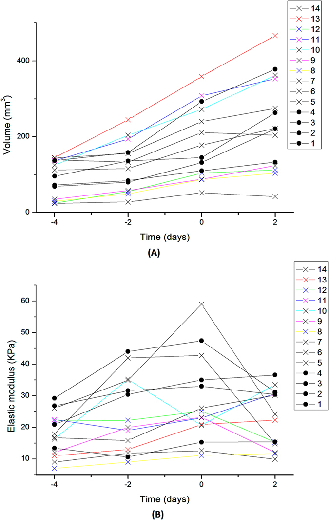
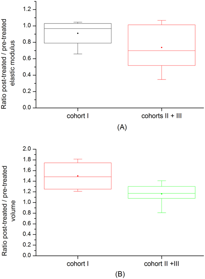
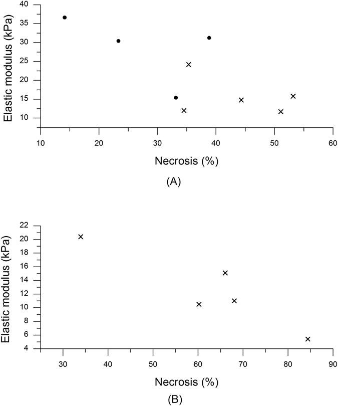
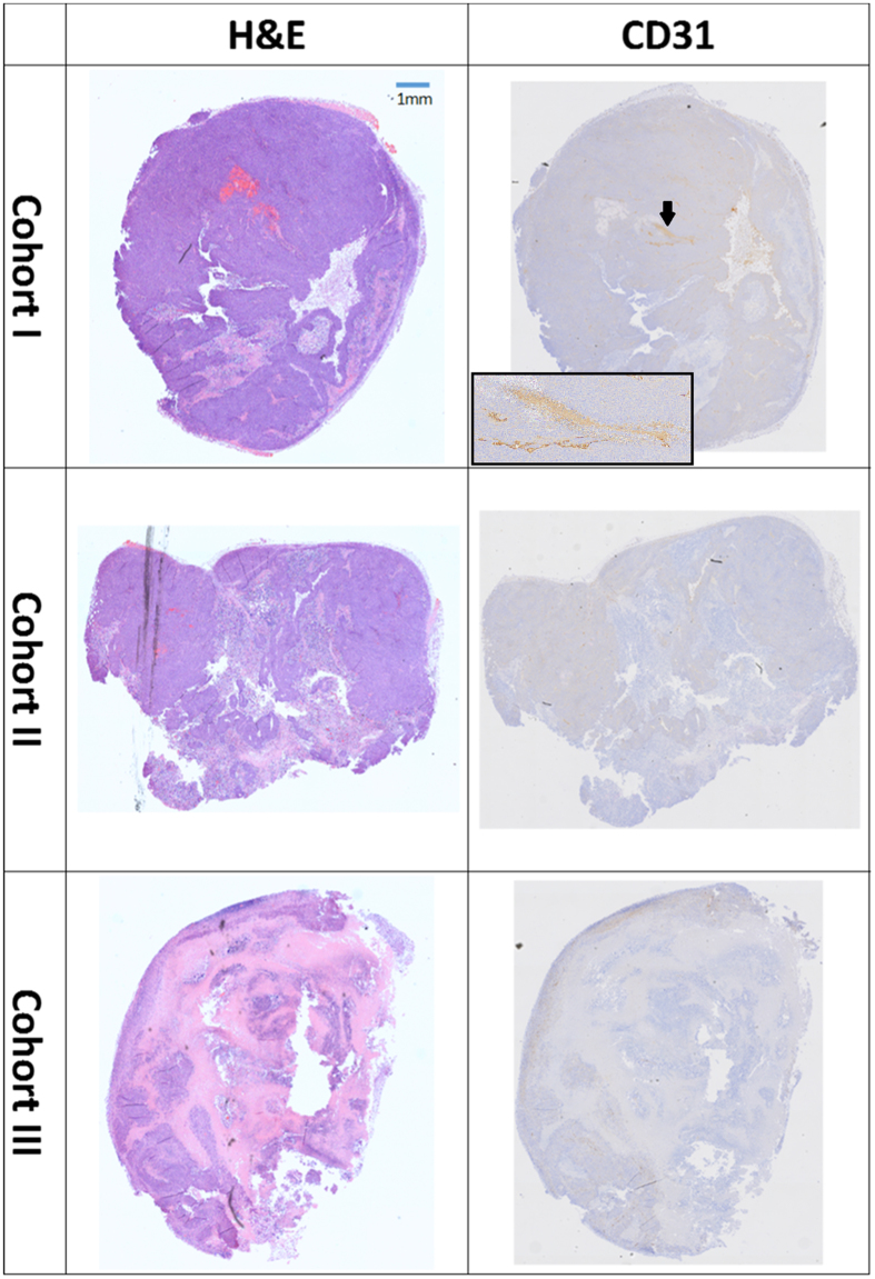
The objective of this study was to evaluate the potential value of ultrasound (US) shear wave elastography (SWE) in assessing the relative change in elastic modulus in colorectal adenocarcinoma xenograft models in vivo and investigate any correlation with histological analysis. We sought to test whether non-invasive evaluation of tissue stiffness is indicative of pathological tumour changes and can be used to monitor therapeutic efficacy. US-SWE was performed in tumour xenografts in 15 NCr nude immunodeficient mice, which were treated with either the cytotoxic drug, Irinotecan, or saline as control. Ten tumours were imaged 48 hours post-treatment and five tumours were imaged for up to five times after treatment. All tumours were harvested for histological analysis and comparison with elasticity measurements. Elastic (Young's) modulus prior to treatment was correlated with tumour volume (r = 0.37, p = 0.008). Irinotecan administration caused significant delay in the tumour growth (p = 0.02) when compared to control, but no significant difference in elastic modulus was detected. Histological analysis revealed a significant correlation between tumour necrosis and elastic modulus (r = -0.73, p = 0.026). SWE measurement provided complimentary information to other imaging modalities and could indicate potential changes in the mechanical properties of tumours, which in turn could be related to the stages of tumour development.
本研究旨在评估超声剪切波弹性成像(SWE)在评估结直肠腺癌异种移植模型体内弹性模量相对变化中的潜在价值,并探讨与组织学分析的相关性。我们旨在测试组织硬度的无创评估是否能指示病理性肿瘤变化,并可用于监测治疗效果。在 15 只 NCr 裸免疫缺陷小鼠的肿瘤异种移植中进行了 US-SWE,其中一些用细胞毒性药物伊立替康治疗,另一些用生理盐水作为对照。在治疗后 48 小时对 10 个肿瘤进行成像,在治疗后最多进行 5 次对 5 个肿瘤进行成像。所有肿瘤均进行了组织学分析,并与弹性测量值进行了比较。治疗前的弹性(杨氏)模量与肿瘤体积呈正相关(r=0.37,p=0.008)。与对照组相比,伊立替康治疗显著延迟了肿瘤生长(p=0.02),但弹性模量无显著差异。组织学分析显示肿瘤坏死与弹性模量之间存在显著相关性(r=-0.73,p=0.026)。SWE 测量提供了与其他成像方式互补的信息,可指示肿瘤力学性质的潜在变化,而这些变化可能与肿瘤发展阶段有关。