Huang Pin-Chieh, Chaney Eric J, Aksamitiene Edita, Barkalifa Ronit, Spillman Darold R, Bogan Bethany J, Boppart Stephen A
Beckman Institute for Advanced Science and Technology, University of Illinois at Urbana-Champaign, USA.
Department of Bioengineering, University of Illinois at Urbana-Champaign, USA.
Theranostics. 2021 Mar 23;11(12):5620-5633. doi: 10.7150/thno.55333. eCollection 2021.
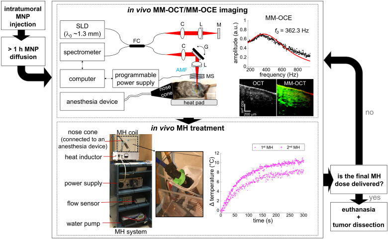
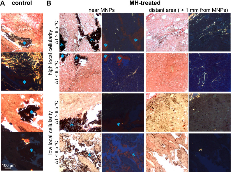
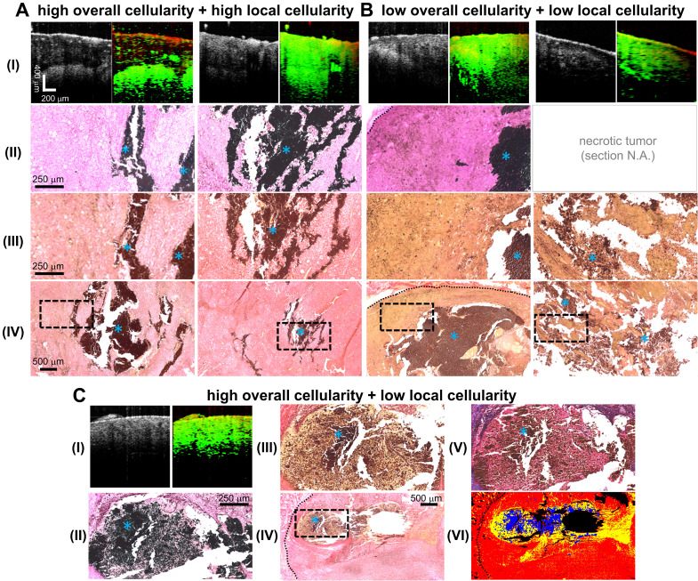
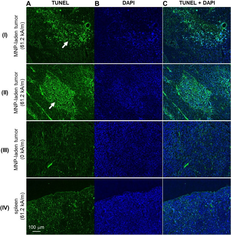
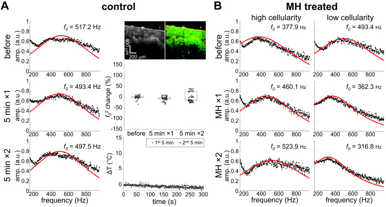
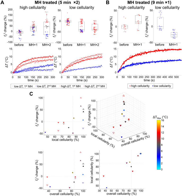
Magnetic nanoparticle hyperthermia (MH) therapy is capable of thermally damaging tumor cells, yet a biomechanically-sensitive monitoring method for the applied thermal dosage has not been established. Biomechanical changes to tissue are known indicators for tumor diagnosis due to its association with the structural organization and composition of tissues at the cellular and molecular level. Here, by exploiting the theranostic functionality of magnetic nanoparticles (MNPs), we aim to explore the potential of using stiffness-based metrics that reveal the intrinsic biophysical changes of melanoma tumors after MH therapy. A total of 14 melanoma-bearing mice were intratumorally injected with dextran-coated MNPs, enabling MH treatment upon the application of an alternating magnetic field (AMF) at 64.7 kHz. The presence of the MNP heating sources was detected by magnetomotive optical coherence tomography (MM-OCT). For the first time, the elasticity alterations of the hyperthermia-treated, MNP-laden, tumors were also measured with magnetomotive optical coherence elastography (MM-OCE), based on the mechanical resonant frequency detected. To investigate the correlation between stiffness changes and the intrinsic biological changes, histopathology was performed on the excised tumor after the measurements. Distinct shifts in mechanical resonant frequency were observed only in the MH-treated group, suggesting a heat-induced stiffness change in the melanoma tumor. Moreover, tumor cellularity, protein conformation, and temperature rise all play a role in tumor stiffness changes after MH treatment. With low cellularity, tumor softens after MH even with low temperature elevation. In contrast, with high cellularity, tumor softening occurs only with a low temperature rise, which is potentially due to protein unfolding, whereas tumor stiffening was seen with a higher temperature rise, likely due to protein denaturation. This study exploits the theranostic functionality of MNPs and investigates the MH-induced stiffness change on melanoma-bearing mice with MM-OCT and MM-OCE for the first time. It was discovered that the elasticity alteration of the melanoma tumor after MH treatment depends on both thermal dosage and the morphological features of the tumor. In summary, changes in tissue-level elasticity can potentially be a physically and physiologically meaningful metric and integrative therapeutic marker for MH treatment, while MM-OCE can be a suitable dosimetry technique.
磁性纳米颗粒热疗(MH)能够对肿瘤细胞造成热损伤,但尚未建立一种对施加的热剂量具有生物力学敏感性的监测方法。由于组织的生物力学变化与细胞和分子水平上组织的结构组织和组成相关联,因此它是肿瘤诊断的已知指标。在此,通过利用磁性纳米颗粒(MNP)的诊疗功能,我们旨在探索使用基于刚度的指标来揭示MH治疗后黑色素瘤肿瘤内在生物物理变化的潜力。总共14只荷黑色素瘤小鼠被瘤内注射了葡聚糖包被的MNP,在施加64.7kHz的交变磁场(AMF)时可进行MH治疗。通过磁动力光学相干断层扫描(MM-OCT)检测MNP热源的存在。首次基于检测到的机械共振频率,用磁动力光学相干弹性成像(MM-OCE)测量了经热疗处理、负载MNP的肿瘤的弹性变化。为了研究刚度变化与内在生物学变化之间的相关性,在测量后对切除的肿瘤进行了组织病理学检查。仅在MH治疗组中观察到机械共振频率的明显变化,表明黑色素瘤肿瘤中存在热诱导的刚度变化。此外,肿瘤细胞密度、蛋白质构象和温度升高均在MH治疗后肿瘤刚度变化中起作用。细胞密度低时,即使温度升高较低,MH治疗后肿瘤也会变软。相反,细胞密度高时,只有在温度升高较低时肿瘤才会变软,这可能是由于蛋白质展开,而温度升高较高时则会出现肿瘤变硬,可能是由于蛋白质变性。本研究利用MNP的诊疗功能,首次用MM-OCT和MM-OCE研究了荷黑色素瘤小鼠MH诱导的刚度变化。发现MH治疗后黑色素瘤肿瘤的弹性变化取决于热剂量和肿瘤的形态特征。总之,组织水平弹性的变化可能是MH治疗的一种物理和生理上有意义的指标及综合治疗标志物,而MM-OCE可能是一种合适的剂量测定技术。