Staples Miranda C, Fannon McKenzie J, Mysore Karthik K, Dutta Rahul R, Ongjoco Alexandria T, Quach Leon W, Kharidia Khush M, Somkuwar Sucharita S, Mandyam Chitra D
Veterans Medical Research Foundation, VA San Diego Healthcare System, La Jolla, CA, USA.
Veterans Medical Research Foundation, VA San Diego Healthcare System, La Jolla, CA, USA.
Brain Res. 2017 May 15;1663:59-65. doi: 10.1016/j.brainres.2017.02.028. Epub 2017 Mar 8.
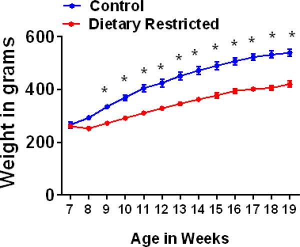
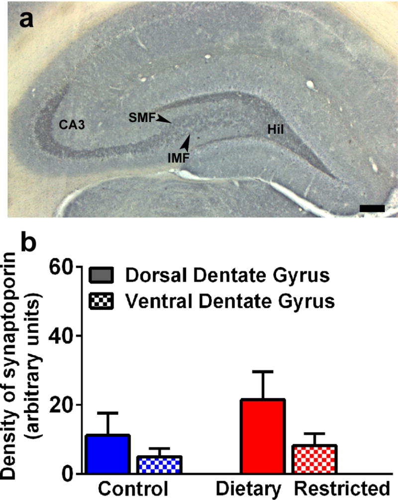
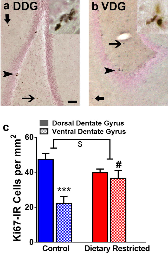
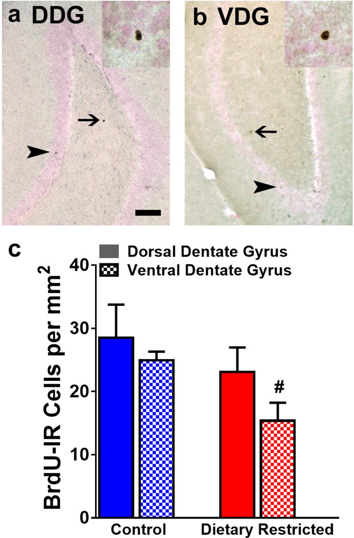
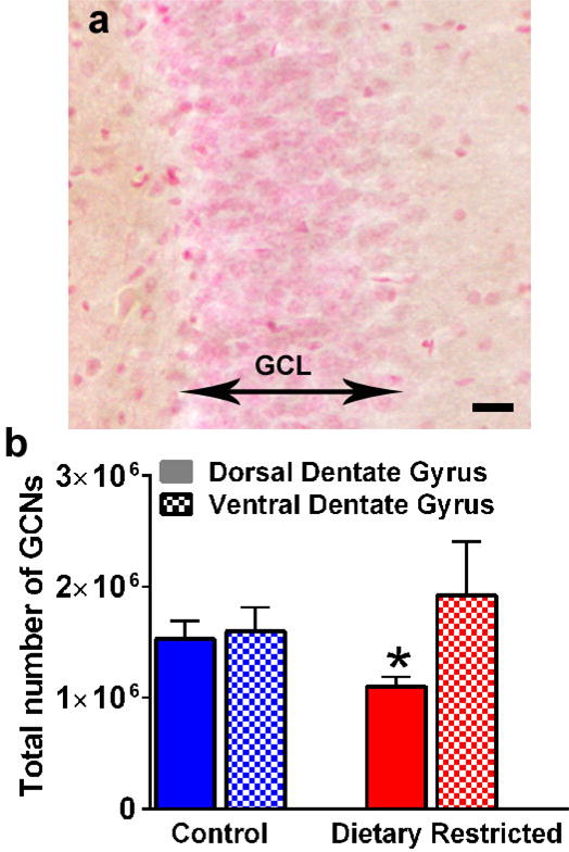
The hippocampal formation undergoes significant morphological and functional changes after prolonged caloric and dietary restriction (DR). In this study we tested whether prolonged DR results in deleterious alterations in hippocampal neurogenesis, density of granule cell neurons and mossy fibers, all of which support plasticity in the dentate gyrus. Young adult animals either experienced free access to food (control condition), or every-other-day feeding regimen (DR condition) for 3months. The number of Ki-67 cells and 28-day old 5-bromo-2'-deoxyuridine (BrdU) cells were quantified in the dorsal and ventral dentate gyrus to determine the effect of DR on cellular proliferation and survival of neural progenitor cells in the anatomically defined regions of the dentate gyrus. The density of granule cell neurons and synaptoporin were also quantified to determine the effect of DR on granule cell neurons and mossy fiber projections in the dentate gyrus. Our results show that DR increases cellular proliferation and concurrently reduces survival of newly born neurons in the ventral dentate gyrus without effecting the number of cells in the dorsal dentate gyrus. DR reduced density of granule cell neurons in the dorsal dentate gyrus. These alterations in the number of granule cell neurons did not affect mossy fiber density in DR animals, which was visualized as no differences in synaptoporin expression. Our findings demonstrate that granule cell neurons in the dentate gyrus are vulnerable to chronic DR and that the reorganization of granule cells in the dentate gyrus subregions is not producing concomitant alterations in dentate gyrus neuronal circuitry with this type of DR.
长期热量限制和饮食限制(DR)后,海马结构会发生显著的形态和功能变化。在本研究中,我们测试了长期DR是否会导致海马神经发生、颗粒细胞神经元密度和苔藓纤维出现有害改变,所有这些都支持齿状回的可塑性。年轻成年动物要么可以自由进食(对照条件),要么采用隔日喂食方案(DR条件),持续3个月。对背侧和腹侧齿状回中Ki-67细胞和28日龄5-溴-2'-脱氧尿苷(BrdU)细胞的数量进行定量,以确定DR对齿状回解剖学定义区域中神经祖细胞的细胞增殖和存活的影响。还对颗粒细胞神经元和突触素的密度进行了定量,以确定DR对齿状回中颗粒细胞神经元和苔藓纤维投射的影响。我们的结果表明,DR增加了腹侧齿状回中的细胞增殖,同时降低了新生神经元的存活率,而对背侧齿状回中的细胞数量没有影响。DR降低了背侧齿状回中颗粒细胞神经元的密度。颗粒细胞神经元数量的这些改变并未影响DR动物中苔藓纤维的密度,这表现为突触素表达没有差异。我们的研究结果表明,齿状回中的颗粒细胞神经元易受慢性DR的影响,并且齿状回亚区域中颗粒细胞的重组并未因这种类型的DR而在齿状回神经元回路中产生相应的改变。