Wu Tao, Hadjantonakis Anna-Katerina, Nowotschin Sonja
Developmental Biology Program, Sloan Kettering Institute, Memorial Sloan Kettering Cancer Center, 1275 York Avenue, New York, NY 10065, USA.
Developmental Biology Program, Sloan Kettering Institute, Memorial Sloan Kettering Cancer Center, 1275 York Avenue, New York, NY 10065, USA
Biol Open. 2017 May 15;6(5):678-687. doi: 10.1242/bio.024638.
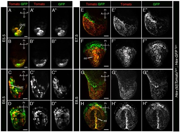
Live imaging is the requisite tool for studying cell behaviors driving embryonic development and tissue formation. Genetically encoded reporters expressed under cell type-specific -regulatory elements that drive fluorescent protein expression at sufficient levels for visualization in living specimens have become indispensable for these studies. Increasingly dual-color (red-green) imaging is used for studying the coordinate behaviors of two cell populations of interest, identifying and characterizing subsets within broader cell populations or subcellular features. Many reporters have been generated using green fluorescent protein (GFP) due to its brightness and developmental neutrality. To compliment the large cohort of available GFP reporters that label cellular populations in early mouse embryos, we have generated a red fluorescent protein (RFP)-based transgenic reporter using the red fluorescent tdTomato protein driven by -regulatory elements from the mouse locus. The reporter predominantly labels endodermal cells. It is a bright RFP-based reporter of the distal visceral endoderm (DVE)/anterior visceral endoderm (AVE), a migratory population within the early post-implantation embryo. It also labels cells of the definitive endoderm (DE), which emerges at gastrulation. Dual-color visualization of these different early endodermal populations will provide a detailed understanding of the cellular behaviors driving key morphogenetic events involving the endoderm.
活体成像技术是研究驱动胚胎发育和组织形成的细胞行为所必需的工具。在细胞类型特异性调控元件控制下表达的遗传编码报告基因,能够驱动荧光蛋白表达至足以在活体标本中进行可视化的水平,对于这些研究来说已经不可或缺。越来越多的双色(红-绿)成像技术被用于研究两个感兴趣的细胞群体的协同行为,识别和表征更广泛细胞群体或亚细胞特征中的子集。由于绿色荧光蛋白(GFP)的亮度和发育中性,许多报告基因都是利用它构建的。为了补充大量可用于标记早期小鼠胚胎细胞群体的GFP报告基因,我们利用小鼠基因座的调控元件驱动红色荧光tdTomato蛋白,构建了一种基于红色荧光蛋白(RFP)的转基因报告基因。该报告基因主要标记内胚层细胞。它是一种基于RFP的明亮报告基因,用于标记远端内脏内胚层(DVE)/前内脏内胚层(AVE),这是植入后早期胚胎中的一个迁移性细胞群体。它还标记原肠胚形成时出现的定形内胚层(DE)细胞。对这些不同的早期内胚层细胞群体进行双色可视化,将有助于详细了解驱动涉及内胚层的关键形态发生事件的细胞行为。