Singh Amandeep, Thukral Chuni Lal, Gupta Kamlesh, Sood Arvinder Singh, Singla Hanish, Singh Kunwarpal
Department of Radiodiagnosis and Imaging, Sri Guru Ramdas Institute of Medical Sciences and Research, Vallah, Amritsar, Punjab, India.
Department of Otorhinolaryngology (ENT), Sri Guru Ramdas Institute of Medical Sciences and Research, Vallah, Amritsar, Punjab, India.
Pol J Radiol. 2017 Feb 16;82:92-99. doi: 10.12659/PJR.899352. eCollection 2017.
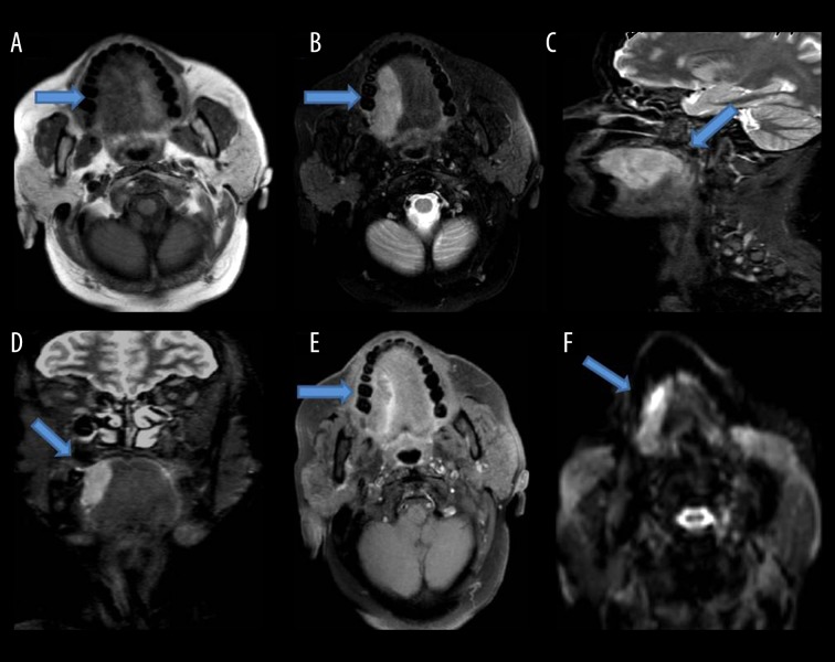
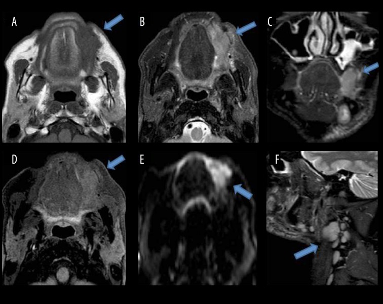
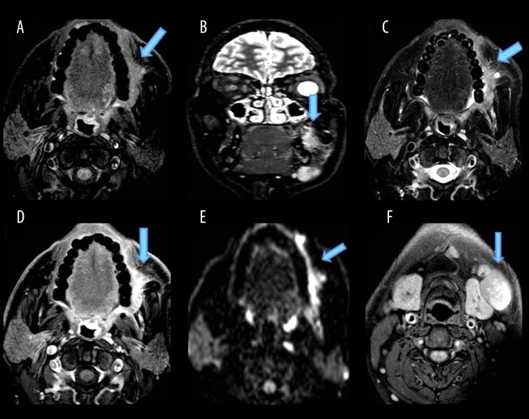
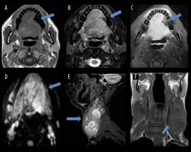
Aim of the present study was to evaluate the role of MRI in staging of malignant lesions of the oral cavity and to correlate MRI findings with clinical/surgical and anatomical-pathological findings, wherever possible.
MATERIAL/METHODS: The study included 50 patients who presented with malignant lesions of the oral cavity and were referred to radiology departments for MRI. All patients included were subjected to a detailed physical examination following which MRI was carried out on Philips Gyroscan Achieva 1.5 Tesla unit.
In the study, the highest number of patients were found to have tongue malignancy (82%) followed by buccal mucosa and gingivobuccal sulcus malignancy (18%). The highest number of patients was in the age group of 51-60 years (32%). The incidence was higher in males (96%). There was moderate agreement (k=0.537) for T stage between the clinical and MRI staging assessments. The agreement for N stage between clinical and MRI staging assessments was fair (k=0.328). The final diagnosis was made by histopathology in 22 patients. The agreement for T stage was good/substantial (k=0.790) and for N stage was moderate (k=0.458) between MRI and histopathology staging assessments.
MRI provides satisfactory accuracy for preoperative estimation of tumor thickness and predicting occult cervical nodal metastasis. MRI is the preferred modality in evaluation and staging of oral cavity malignancy which helps a clinician for planning of treatment.
本研究的目的是评估MRI在口腔恶性病变分期中的作用,并尽可能将MRI表现与临床/手术及解剖病理学表现进行关联。
材料/方法:该研究纳入了50例患有口腔恶性病变并被转诊至放射科进行MRI检查的患者。所有纳入的患者均接受了详细的体格检查,之后在飞利浦Gyroscan Achieva 1.5特斯拉设备上进行了MRI检查。
在该研究中,发现舌部恶性肿瘤患者数量最多(82%),其次是颊黏膜和龈颊沟恶性肿瘤(18%)。患者数量最多的年龄组为51 - 60岁(32%)。男性发病率较高(96%)。临床和MRI分期评估之间T分期的一致性为中等(k = 0.537)。临床和MRI分期评估之间N分期的一致性为一般(k = 0.328)。22例患者通过组织病理学做出最终诊断。MRI和组织病理学分期评估之间T分期的一致性良好/高度一致(k = 0.790),N分期的一致性为中等(k = 0.458)。
MRI在术前评估肿瘤厚度和预测隐匿性颈部淋巴结转移方面具有令人满意的准确性。MRI是评估口腔恶性肿瘤及其分期的首选方式,有助于临床医生制定治疗方案。