Olson Elizabeth A, Cui Jiaolong, Fukunaga Rena, Nickerson Lisa D, Rauch Scott L, Rosso Isabelle M
Center for Depression, Anxiety and Stress Research, McLean Hospital, Belmont, MA, USA.
Department of Psychiatry, Harvard Medical School, Boston, MA, USA.
Depress Anxiety. 2017 May;34(5):437-445. doi: 10.1002/da.22615. Epub 2017 Mar 15.
Most studies of brain white matter (WM) in posttraumatic stress disorder (PTSD) have focused on combat trauma, and often were confounded by neurological and substance dependence comorbidity. This study used tract-based spatial statistics (TBSS) and probabilistic tractography to characterize WM microstructure in a mixed-sex community sample of PTSD patients exposed to diverse and multiple traumas, and in trauma-exposed normal comparison (TENC) subjects.
TBSS compared diffusion measures between 20 adults with DSM-IV PTSD and 17 TENC, using a whole-brain voxel-wise approach. Probabilistic tractography using Freesurfer's TRACULA was employed to measure diffusion tensor imaging (DTI) metrics within anatomically defined pathways. DTI metrics were compared between groups and correlated with PTSD symptom severity and trauma load.
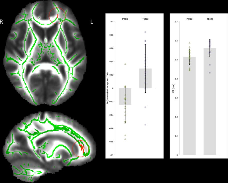
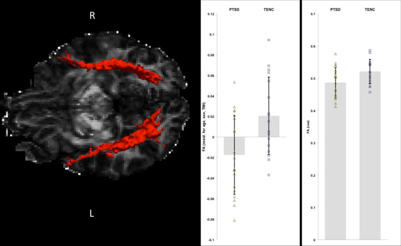
Controlling for age, sex, and motion, PTSD subjects had significantly reduced fractional anisotropy (FA) in a left frontal lobe cluster compared with TENC, at p < .05, family-wise error corrected. Tractography identified significant group differences in the inferior longitudinal fasciculus (ILF), including lower FA and higher radial diffusivity in PTSD compared with TENC. Within the PTSD group, FA values were not correlated with symptom severity or trauma load. Results remained significant after removing participants using psychotropic medication or those with comorbid major depression.
PTSD patients had reduced WM integrity in left hemisphere frontal WM and temporal-occipital WM tracts, compared to trauma-exposed controls. Reduced frontal FA is consistent with compromised top-down attentional control and emotion regulation in PTSD, while reduced ILF FA may be related to sensory processing and gating abnormalities in this disorder.
大多数关于创伤后应激障碍(PTSD)患者脑白质(WM)的研究都集中在战斗创伤上,并且常常受到神经学和物质依赖共病的干扰。本研究采用基于体素的空间统计学(TBSS)和概率纤维束成像技术,对暴露于多种不同创伤的PTSD患者的混合性别社区样本以及暴露于创伤的正常对照(TENC)受试者的WM微观结构进行表征。
TBSS采用全脑体素水平的方法,比较了20名符合《精神疾病诊断与统计手册》第四版(DSM-IV)PTSD诊断标准的成年人与17名TENC受试者之间的扩散指标。使用Freesurfer的TRACULA进行概率纤维束成像,以测量解剖学定义通路内的扩散张量成像(DTI)指标。比较两组之间的DTI指标,并将其与PTSD症状严重程度和创伤负荷相关联。
在控制年龄、性别和运动因素后,与TENC相比,PTSD受试者左侧额叶簇的分数各向异性(FA)显著降低,经家族性错误校正后,p < 0.05。纤维束成像显示,在颞下纵束(ILF)中存在显著的组间差异,与TENC相比,PTSD患者的FA值较低,径向扩散率较高。在PTSD组内,FA值与症状严重程度或创伤负荷无关。在排除使用精神药物的参与者或患有共病重度抑郁症的参与者后,结果仍然显著。
与暴露于创伤的对照组相比,PTSD患者左半球额叶WM和颞枕叶WM束的WM完整性降低。额叶FA降低与PTSD中自上而下的注意力控制和情绪调节受损一致,而ILF FA降低可能与该疾病中的感觉处理和门控异常有关。