From the Department of Neuroscience, Science for Life Laboratory, Uppsala University, Sweden (Gu, Dubol, Comasco); the Department of Clinical Sciences, Umeå University, Umeå, Sweden (Stiernman, Bixo); the Department of Surgical Sciences, Neuroradiology, Uppsala University, Sweden (Wikström); the Department of Psychiatry and Psychotherapy, Medical University of Vienna, Austria (Hahn, Lanzenberger); the Department of Psychiatry, University of Colorado School of Medicine, USA (Epperson); the Department of Women's and Children's Health, Uppsala University, Sweden (Sundström-Poromaa).
From the Department of Neuroscience, Science for Life Laboratory, Uppsala University, Sweden (Gu, Dubol, Comasco); the Department of Clinical Sciences, Umeå University, Umeå, Sweden (Stiernman, Bixo); the Department of Surgical Sciences, Neuroradiology, Uppsala University, Sweden (Wikström); the Department of Psychiatry and Psychotherapy, Medical University of Vienna, Austria (Hahn, Lanzenberger); the Department of Psychiatry, University of Colorado School of Medicine, USA (Epperson); the Department of Women's and Children's Health, Uppsala University, Sweden (Sundström-Poromaa)
J Psychiatry Neurosci. 2022 Feb 23;47(1):E67-E76. doi: 10.1503/jpn.210143. Print 2022 Jan-Feb.
Premenstrual dysphoric disorder (PMDD) is a mood disorder characterized by psychological and physical symptoms. Differences in white matter have been associated with affective and anxiety disorders, which share some symptoms with PMDD. However, whether white matter structure differs between the brains of individuals with PMDD and healthy controls is not known, nor is its relation to symptom severity.
We performed tract-based spatial statistics and voxel-based morphometry analyses of diffusion tensor imaging metrics and white matter volume, using 2 neuroimaging data sets ( = 67 and = 131) and a combined whole-brain and region-of-interest approach. We performed correlation analyses to investigate the relationship between regions with different white matter microstructure and volume and PMDD symptom severity.
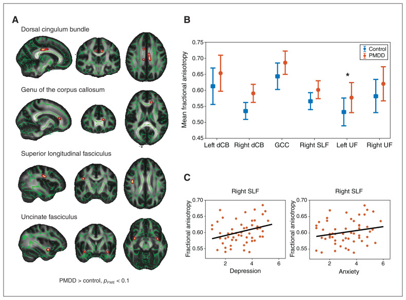
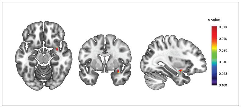
We found greater fractional anisotropy in the left uncinate fasciculus ( = 0.69) in individuals with PMDD compared to controls. Moreover, the volume of the right uncinate fasciculus was higher in individuals with PMDD compared to controls ( = 0.40). As well, the severity of premenstrual depression was positively correlated with fractional anisotropy in the right superior longitudinal fasciculus ( = 0.35).
It is challenging to interpret group differences in diffusion tensor imaging metrics in terms of their underlying biophysical properties. The small size of the control group in the diffusion tensor imaging study may have prevented effects of interest from being detected.
The findings of the present study provide evidence of differential cerebral white matter structure associated with PMDD and its symptoms.
经前烦躁障碍(PMDD)是一种以心理和生理症状为特征的情绪障碍。已有研究表明,大脑白质的差异与情感障碍和焦虑障碍有关,而这些障碍与 PMDD 有一些共同的症状。然而,PMDD 患者与健康对照组的大脑白质结构是否存在差异,以及其与症状严重程度的关系,目前尚不清楚。
我们使用 2 个神经影像学数据集(= 67 和= 131)和全脑及感兴趣区的联合方法,进行了基于束的空间统计学和基于体素的形态计量学分析,以研究扩散张量成像指标和白质体积的白质结构。我们进行了相关性分析,以研究具有不同白质微观结构和体积的区域与 PMDD 症状严重程度之间的关系。
与对照组相比,PMDD 患者左侧钩束的各向异性分数更高(= 0.69)。此外,PMDD 患者右侧钩束的体积高于对照组(= 0.40)。而且,经前抑郁的严重程度与右侧上纵束的各向异性分数呈正相关(= 0.35)。
根据扩散张量成像指标的潜在生物物理特性来解释组间差异具有一定的挑战性。扩散张量成像研究中对照组的规模较小,可能导致感兴趣的效应无法被检测到。
本研究的结果提供了与 PMDD 及其症状相关的大脑白质结构差异的证据。