Siehl Sebastian, Wicking Manon, Pohlack Sebastian, Winkelmann Tobias, Zidda Francesca, Steiger-White Frauke, King John, Burgess Neil, Flor Herta, Nees Frauke
Institute of Cognitive and Clinical Neuroscience, Central Institute of Mental Health, Medical Faculty Mannheim, Ruprecht-Karls-University Heidelberg, Mannheim, Germany; Graduate School of Economic and Social Sciences, University of Mannheim, Mannheim, Germany; UCL Institute of Cognitive Neuroscience, University College London, London, United Kingdom.
Institute of Cognitive and Clinical Neuroscience, Central Institute of Mental Health, Medical Faculty Mannheim, Ruprecht-Karls-University Heidelberg, Mannheim, Germany; Department of Pain Medicine, BG University Hospital Bergmannsheil GmbH, Ruhr University, Bochum, Germany.
Neuroimage Clin. 2020;28:102424. doi: 10.1016/j.nicl.2020.102424. Epub 2020 Sep 12.
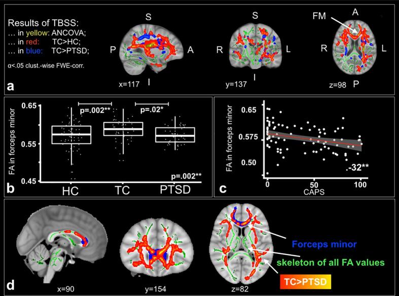
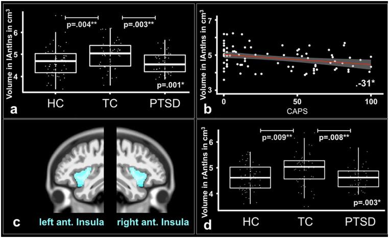
Differences in structural white and gray matter in survivors of traumatic experiences have been related to the development and maintenance of Posttraumatic Stress Disorder (PTSD). However, there are very few studies on diffusion tensor imaging and region based morphometry comparing patients with PTSD to two control groups, namely healthy individuals with or without trauma experience. It is also unknown if differences in white and gray matter are associated. In this cross-sectional study, we examined white- and gray matter differences between 44 patients with PTSD, 49 trauma control and 61 healthy control subjects. We compared the groups applying Tract-Based Spatial Statistics (TBSS) for a whole brain white matter analysis as well as region of interest analyses for white and gray matter. First, trauma control subjects in comparison to patients with PTSD and healthy control subjects showed significantly a) higher fractional anisotropy (FA) in the left corticospinal tract and inferior fronto-occipital fasciculus than patients with PTSD, b) higher FA in the left inferior fronto-occipital-, right inferior- and right superior longitudinal fasciculi, c) higher FA in the forceps minor and d) higher volume of the left and right anterior insulae. Second, we show significant correlations between the FA in the forceps minor and the gray matter volume in the left and right anterior insulae. Third, the mean FA value in the forceps minor correlated negatively with symptom severity of PTSD and depression as well as trait anxiety, whereas the gray matter volume in the left anterior insula correlated negatively with symptom severity in PTSD. Our findings underline the importance of brain structures critically involved in emotion regulation and salience mapping. While previous studies associated these processes primarily to functional and task-based differences in brain activity, we argue that morphometrical white and gray matter differences could serve as targets in neuroscientifically-informed prevention and treatment interventions for PTSD.
创伤经历幸存者的白质和灰质结构差异与创伤后应激障碍(PTSD)的发生和维持有关。然而,关于扩散张量成像和基于区域的形态测量学,将PTSD患者与两个对照组(即有或没有创伤经历的健康个体)进行比较的研究非常少。白质和灰质的差异是否相关也尚不清楚。在这项横断面研究中,我们检查了44例PTSD患者、49例创伤对照组和61例健康对照受试者之间的白质和灰质差异。我们应用基于体素的空间统计学(TBSS)对全脑白质进行分析,并对白质和灰质进行感兴趣区域分析,以比较各组。首先,与PTSD患者和健康对照受试者相比,创伤对照组受试者在以下方面表现出显著差异:a)左侧皮质脊髓束和额枕下束的分数各向异性(FA)高于PTSD患者;b)左侧额枕下束、右侧下纵束和右侧上纵束的FA较高;c)小钳夹的FA较高;d)左右前岛叶的体积较大。其次,我们发现小钳夹的FA与左右前岛叶的灰质体积之间存在显著相关性。第三,小钳夹的平均FA值与PTSD、抑郁症状严重程度以及特质焦虑呈负相关,而左侧前岛叶的灰质体积与PTSD症状严重程度呈负相关。我们的研究结果强调了大脑结构在情绪调节和显著性映射中所起的关键作用。虽然先前的研究主要将这些过程与大脑活动中的功能和基于任务的差异联系起来,但我们认为形态学上的白质和灰质差异可以作为PTSD神经科学导向预防和治疗干预的靶点。