• 文献检索
  • 文档翻译
  • 深度研究
  • 学术资讯
  • Suppr Zotero 插件Zotero 插件
  • 邀请有礼
  • 套餐&价格
  • 历史记录
应用&插件
Suppr Zotero 插件Zotero 插件浏览器插件Mac 客户端Windows 客户端微信小程序
定价
高级版会员购买积分包购买API积分包
服务
文献检索文档翻译深度研究API 文档MCP 服务
关于我们
关于 Suppr公司介绍联系我们用户协议隐私条款
关注我们

Suppr 超能文献

核心技术专利:CN118964589B侵权必究
粤ICP备2023148730 号-1Suppr @ 2026

文献检索

告别复杂PubMed语法,用中文像聊天一样搜索,搜遍4000万医学文献。AI智能推荐,让科研检索更轻松。

立即免费搜索

文件翻译

保留排版,准确专业,支持PDF/Word/PPT等文件格式,支持 12+语言互译。

免费翻译文档

深度研究

AI帮你快速写综述,25分钟生成高质量综述,智能提取关键信息,辅助科研写作。

立即免费体验

倾斜或拉伸光学相干断层扫描图像中视网膜厚度测量的变异性

Variability of Retinal Thickness Measurements in Tilted or Stretched Optical Coherence Tomography Images.

作者信息

Uji Akihito, Abdelfattah Nizar Saleh, Boyer David S, Balasubramanian Siva, Lei Jianqin, Sadda SriniVas R

机构信息

Doheny Image Reading Center, Doheny Eye Institute, Los Angeles, California, USA ; Department of Ophthalmology, David Geffen School of Medicine at the University of California-Los Angeles, Los Angeles, California, USA.

Retina Vitreous Associates Medical Group, Beverly Hills, California, USA.

出版信息

Transl Vis Sci Technol. 2017 Mar 1;6(2):1. doi: 10.1167/tvst.6.2.1. eCollection 2017 Mar.

DOI:10.1167/tvst.6.2.1
PMID:28299239
原文链接:https://pmc.ncbi.nlm.nih.gov/articles/PMC5347660/
Abstract

PURPOSE

To investigate the level of inaccuracy of retinal thickness measurements in tilted and axially stretched optical coherence tomography (OCT) images.

METHODS

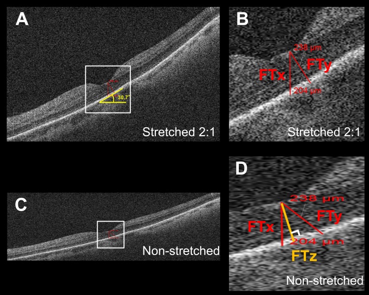
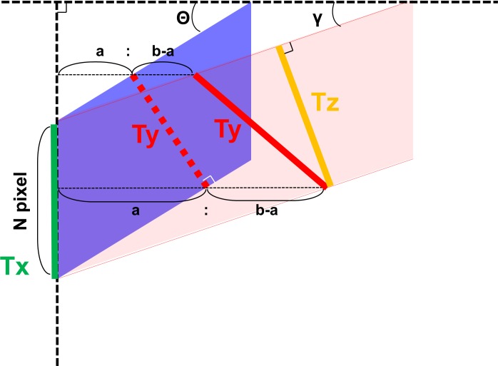
A consecutive series of 50 eyes of 50 patients with age-related macular degeneration were included in this study, and Cirrus HD-OCT images through the foveal center were used for the analysis. The foveal thickness was measured in three ways: (1) parallel to the orientation of the A-scan (Tx), (2) perpendicular to the retinal pigment epithelium (RPE) surface in the instrument-displayed aspect ratio image (Ty), and (3) thickness measured perpendicular to the RPE surface in a native aspect ratio image (Tz). Mathematical modeling was performed to estimate the measurement error.

RESULTS

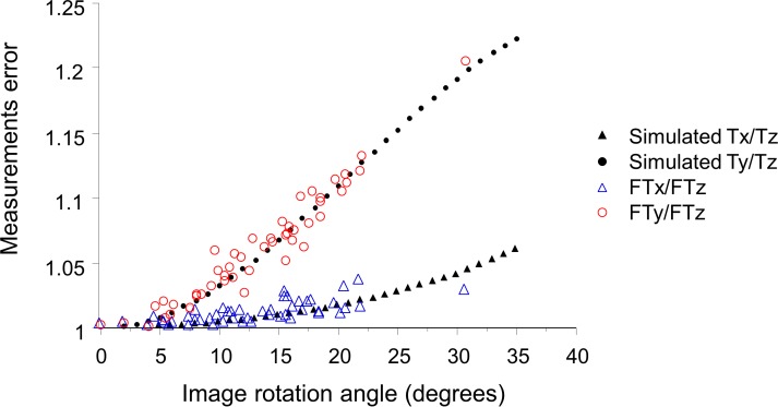
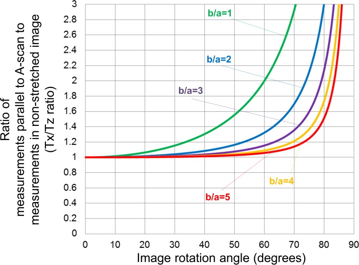
The measurement error was larger in tilted images with a greater angle of tilt. In the simulation, with axial stretching by a factor of 2, Ty/Tz ratio was > 1.05 at a tilt angle between 13° to 18° and 72° to 77°, > 1.10 at a tilt angle between 19° to 31° and 59° to 71°, and > 1.20 at an angle ranging from 32° to 58°. Of note with even more axial stretching, the Ty/Tz ratio is even larger. Tx/Tz ratio was smaller than the Ty/Tz ratio at angles ranging from 0° to 54°. The actual patient data showed good agreement with the simulation.

UNLABELLED

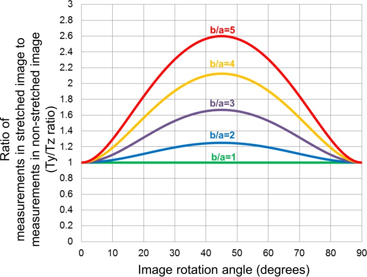
The Ty/Tz ratio was greater than 1.05 (5% error) at angles ranging from 13° to 18° and 72° to 77°, greater than 1.10 (10% error) angles ranging from 19° to 31° and 59° to 71°, and greater than 1.20 (20% error) angles ranging from 32° to 58° in the images axially stretched by a factor of 2 (b/a = 2), which is typical of most OCT instrument displays.

CONCLUSIONS

Retinal thickness measurements obtained perpendicular to the RPE surface were overestimated when using tilted and axially stretched OCT images.

TRANSLATIONAL RELEVANCE

If accurate measurements are to be obtained, images with a native aspect ratio similar to microscopy must be used.

摘要

目的

研究倾斜和轴向拉伸的光学相干断层扫描(OCT)图像中视网膜厚度测量的不准确程度。

方法

本研究纳入了50例年龄相关性黄斑变性患者的连续50只眼,使用通过黄斑中心的Cirrus HD-OCT图像进行分析。通过三种方式测量黄斑厚度:(1)平行于A扫描方向(Tx);(2)在仪器显示的长宽比图像中垂直于视网膜色素上皮(RPE)表面(Ty);(3)在原始长宽比图像中垂直于RPE表面测量的厚度(Tz)。进行数学建模以估计测量误差。

结果

倾斜角度越大的倾斜图像中测量误差越大。在模拟中,轴向拉伸2倍时,在13°至18°和72°至77°的倾斜角度下,Ty/Tz比值>1.05;在19°至31°和59°至71°的倾斜角度下,>1.10;在32°至58°的角度范围内,>1.20。值得注意的是,轴向拉伸越大,Ty/Tz比值越大。在0°至54°的角度范围内,Tx/Tz比值小于Ty/Tz比值。实际患者数据与模拟结果吻合良好。

未标注

在轴向拉伸2倍(b/a = 2)的图像中,这是大多数OCT仪器显示的典型情况,在13°至18°和72°至77°的角度范围内,Ty/Tz比值大于1.05(误差5%);在19°至31°和59°至71°的角度范围内,大于1.10(误差10%);在32°至58°的角度范围内,大于1.20(误差20%)。

结论

使用倾斜和轴向拉伸的OCT图像时,垂直于RPE表面获得的视网膜厚度测量值被高估。

转化相关性

若要获得准确测量值,则必须使用与显微镜相似的原始长宽比图像。

https://cdn.ncbi.nlm.nih.gov/pmc/blobs/262e/5347660/0806573fbe64/i2164-2591-6-2-1-f05.jpg
https://cdn.ncbi.nlm.nih.gov/pmc/blobs/262e/5347660/4f75dbee9069/i2164-2591-6-2-1-f01.jpg
https://cdn.ncbi.nlm.nih.gov/pmc/blobs/262e/5347660/a3fd0a9059f5/i2164-2591-6-2-1-f02.jpg
https://cdn.ncbi.nlm.nih.gov/pmc/blobs/262e/5347660/0dd807a215d5/i2164-2591-6-2-1-f03.jpg
https://cdn.ncbi.nlm.nih.gov/pmc/blobs/262e/5347660/73b70561d054/i2164-2591-6-2-1-f04.jpg
https://cdn.ncbi.nlm.nih.gov/pmc/blobs/262e/5347660/0806573fbe64/i2164-2591-6-2-1-f05.jpg

相似文献

1
Variability of Retinal Thickness Measurements in Tilted or Stretched Optical Coherence Tomography Images.倾斜或拉伸光学相干断层扫描图像中视网膜厚度测量的变异性
Transl Vis Sci Technol. 2017 Mar 1;6(2):1. doi: 10.1167/tvst.6.2.1. eCollection 2017 Mar.
2
Alignment artifacts in optical coherence tomography analyzed images.光学相干断层扫描分析图像中的对准伪影。
Ophthalmology. 2007 Feb;114(2):263-70. doi: 10.1016/j.ophtha.2006.06.059. Epub 2006 Nov 21.
3
Myopic optic disc tilt and the characteristics of peripapillary retinal nerve fiber layer thickness measured by spectral-domain optical coherence tomography.高度近视视盘倾斜与频域光学相干断层扫描测量的视盘周围视网膜神经纤维层厚度的特征。
J Glaucoma. 2012 Apr-May;21(4):260-5. doi: 10.1097/IJG.0b013e31820719e1.
4
Characteristics of peripapillary retinal nerve fiber layer thickness in eyes with myopic optic disc tilt and rotation.近视性视盘倾斜和旋转眼的视盘旁视网膜神经纤维层厚度特征。
J Glaucoma. 2012 Aug;21(6):394-400. doi: 10.1097/IJG.0b013e3182182567.
5
Impact of scanning density on measurements from spectral domain optical coherence tomography.扫描密度对光谱域光学相干断层扫描测量结果的影响。
Invest Ophthalmol Vis Sci. 2010 Feb;51(2):1071-8. doi: 10.1167/iovs.09-4325. Epub 2009 Sep 24.
6
Fully automatic software for retinal thickness in eyes with diabetic macular edema from images acquired by cirrus and spectralis systems.用于从 cirrus 和 spectralis 系统获取的糖尿病性黄斑水肿眼图像进行视网膜厚度全自动分析的软件。
Invest Ophthalmol Vis Sci. 2013 Nov 15;54(12):7595-602. doi: 10.1167/iovs.13-11762.
7
Comparison of spectral-domain versus time-domain optical coherence tomography in management of age-related macular degeneration with ranibizumab.频域与时域光学相干断层扫描在雷珠单抗治疗年龄相关性黄斑变性中的比较
Ophthalmology. 2009 May;116(5):947-55. doi: 10.1016/j.ophtha.2008.11.002. Epub 2009 Feb 20.
8
Prospective comparison of cirrus and stratus optical coherence tomography for quantifying retinal thickness.Cirrus和Stratus光学相干断层扫描技术在定量测量视网膜厚度方面的前瞻性比较。
Am J Ophthalmol. 2009 Feb;147(2):267-275.e2. doi: 10.1016/j.ajo.2008.08.018. Epub 2008 Oct 17.
9
Errors in retinal thickness measurements obtained by optical coherence tomography.光学相干断层扫描获得的视网膜厚度测量误差。
Ophthalmology. 2006 Feb;113(2):285-93. doi: 10.1016/j.ophtha.2005.10.005. Epub 2006 Jan 10.
10
[Fundus features of nanophthalmos analyzed by optical coherence tomography].[光学相干断层扫描分析小眼球的眼底特征]
Zhonghua Yan Ke Za Zhi. 2013 Dec;49(12):1069-74.

引用本文的文献

1
Peripheral retinal and choroidal thickness of eyes with reticular pseudodrusen.伴有网状假性玻璃膜疣的眼睛的周边视网膜和脉络膜厚度
Graefes Arch Clin Exp Ophthalmol. 2025 May 3. doi: 10.1007/s00417-025-06842-5.
2
Derivation of human retinal cell densities using high-density, spatially localized optical coherence tomography data from the human retina.利用高密度、空间定位的人眼视网膜光学相干断层扫描数据推导人视网膜细胞密度。
J Comp Neurol. 2023 Aug;531(11):1108-1125. doi: 10.1002/cne.25483. Epub 2023 Apr 19.
3
Early localized alterations of the retinal inner plexiform layer in association with visual field worsening in glaucoma patients.

本文引用的文献

1
PROGRESSION OF MACULAR ATROPHY IN PATIENTS WITH NEOVASCULAR AGE-RELATED MACULAR DEGENERATION UNDERGOING ANTIVASCULAR ENDOTHELIAL GROWTH FACTOR THERAPY.接受抗血管内皮生长因子治疗的新生血管性年龄相关性黄斑变性患者黄斑萎缩的进展
Retina. 2016 Oct;36(10):1843-50. doi: 10.1097/IAE.0000000000001059.
2
Tissue thickness calculation in ocular optical coherence tomography.眼部光学相干断层扫描中的组织厚度计算。
Biomed Opt Express. 2016 Jan 21;7(2):629-45. doi: 10.1364/BOE.7.000629. eCollection 2016 Feb 1.
3
A Problem of Proportions in OCT-Based Morphometry and a Proposed Solution.
青光眼患者视野恶化相关的视网膜内丛状层早期局限性改变。
PLoS One. 2021 Feb 25;16(2):e0247401. doi: 10.1371/journal.pone.0247401. eCollection 2021.
4
Correlation of spectral domain optical coherence tomography with histology and electron microscopy in the porcine retina.谱域光学相干断层扫描与组织学和电镜在猪眼视网膜的相关性。
Exp Eye Res. 2018 Dec;177:181-190. doi: 10.1016/j.exer.2018.08.003. Epub 2018 Aug 16.
基于光学相干断层扫描的形态测量中的比例问题及一种解决方案
Invest Ophthalmol Vis Sci. 2016 Feb;57(2):484-5. doi: 10.1167/iovs.15-18570.
4
Potential measurement errors due to image enlargement in optical coherence tomography imaging.光学相干断层扫描成像中由于图像放大导致的潜在测量误差。
PLoS One. 2015 May 29;10(5):e0128512. doi: 10.1371/journal.pone.0128512. eCollection 2015.
5
Application of extended field imaging to optical coherence tomography.扩展视野成像在光学相干断层扫描中的应用。
Ophthalmology. 2015 Jun;122(6):1272-4. doi: 10.1016/j.ophtha.2014.12.035. Epub 2015 Jan 29.
6
Influence of axial length on thickness measurements using spectral-domain optical coherence tomography.眼轴长度对使用光谱域光学相干断层扫描进行厚度测量的影响。
Invest Ophthalmol Vis Sci. 2014 Oct 8;55(11):7494-8. doi: 10.1167/iovs.14-14043.
7
Megahertz ultra-wide-field swept-source retina optical coherence tomography compared to current existing imaging devices.与现有成像设备相比的兆赫兹超宽视野扫频源视网膜光学相干断层扫描
Graefes Arch Clin Exp Ophthalmol. 2014 Jun;252(6):1009-16. doi: 10.1007/s00417-014-2640-4. Epub 2014 May 1.
8
Parallelism as a novel marker for structural integrity of retinal layers in optical coherence tomographic images in eyes with epiretinal membrane.在患有视网膜内界膜的眼睛的光学相干断层扫描图像中,平行性作为视网膜层结构完整性的新标志物。
Am J Ophthalmol. 2014 Jan;157(1):227-236.e4. doi: 10.1016/j.ajo.2013.09.008. Epub 2013 Oct 16.
9
Multiple layer segmentation and analysis in three-dimensional spectral-domain optical coherence tomography volume scans.三维频域光学相干断层扫描容积扫描的多层分割与分析。
J Biomed Opt. 2013 Jul;18(7):76006. doi: 10.1117/1.JBO.18.7.076006.
10
Adjustment of the retinal angle in SD-OCT of glaucomatous eyes provides better intervisit reproducibility of peripapillary RNFL thickness.调整青光眼患者的视网膜角度可提高视盘周围视网膜神经纤维层厚度的随访间可重复性。
Invest Ophthalmol Vis Sci. 2013 Jul 18;54(7):4808-12. doi: 10.1167/iovs.13-12211.