Talebi Farideh, Ghorbani Samira, Chan Wing Fuk, Boghozian Roobina, Masoumi Farimah, Ghasemi Sedigheh, Vojgani Mohammed, Power Christopher, Noorbakhsh Farshid
Department of Immunology, School of Medicine, Tehran University of Medical Sciences, Tehran, Iran.
Shefa Neuroscience Research Institute, Khatam Al-Anbia Hospital, Tehran, Iran.
J Neuroinflammation. 2017 Mar 16;14(1):55. doi: 10.1186/s12974-017-0832-7.
MicroRNAs have emerged as an important class of modulators of gene expression. These molecules influence protein synthesis through translational repression or degradation of mRNA transcripts. Herein, we investigated the potential role of miR-142a isoforms, miR-142a-3p and miR-142a-5p, in the context of autoimmune neuroinflammation.
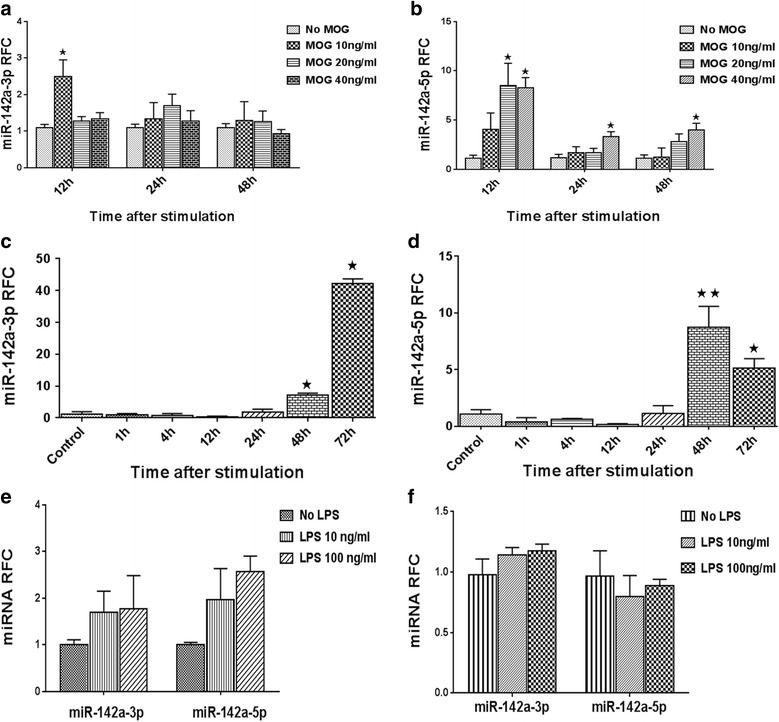
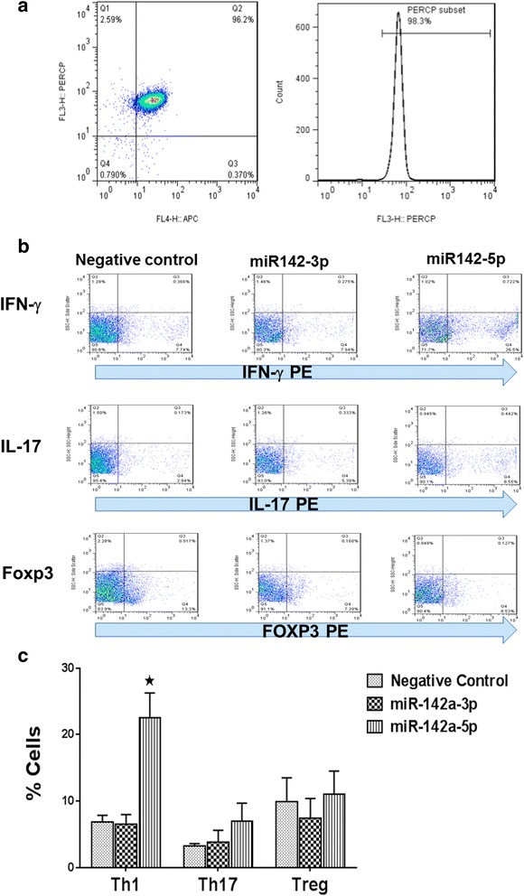
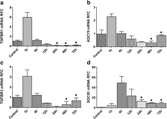
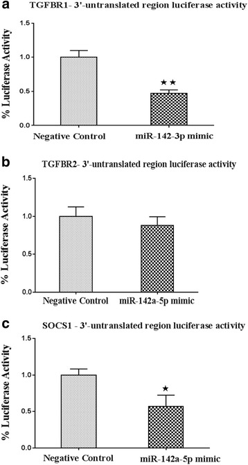
The expression levels of two mature isoforms of miR-142 were measured in the brains of patients with multiple sclerosis (MS) and the CNS tissues from mice with experimental autoimmune encephalomyelitis (EAE), an animal model of MS. Expression analyses were also performed in mitogen and antigen-stimulated splenocytes, as well as macrophages and astrocytes using real-time RT-PCR. The role of the mature miRNAs was then investigated in T cell differentiation by transfection of CD4 T cells, followed by flow cytometric analysis of intracellular cytokines. Luciferase assays using vectors containing the 3'UTR of predicted targets were performed to confirm the interaction of miRNA sequences with transcripts. Expression of targets were then analyzed in activated splenocytes and MS/EAE tissues.
Expression of miR-142-5p was significantly increased in the frontal white matter from MS patients compared with white matter from non-MS controls. Likewise, expression levels of miR-142a-5p and miR-142a-3p showed significant upregulation in the spinal cords of EAE mice at days 15 and 25 post disease induction. Splenocytes stimulated with myelin oligodendrocyte glycoprotein (MOG) peptide or anti-CD3/anti-CD28 antibodies showed upregulation of miR-142a-5p and miR-142a-3p isoforms, whereas stimulated bone marrow-derived macrophages and primary astrocytes did not show any significant changes in miRNA expression levels. miR-142a-5p overexpression in activated lymphocytes shifted the pattern of T cell differentiation towards Th1 cells. Luciferase assays revealed SOCS1 and TGFBR1 as direct targets of miR-142a-5p and miR-142a-3p, respectively, and overexpression of miRNA mimic sequences suppressed the expression of these target transcripts in lymphocytes. SOCS1 levels were also diminished in MS white matter and EAE spinal cords.
Our findings suggest that increased expression of miR-142 isoforms might be involved in the pathogenesis of autoimmune neuroinflammation by influencing T cell differentiation, and this effect could be mediated by interaction of miR-142 isoforms with SOCS1 and TGFBR-1 transcripts.
微小RNA已成为一类重要的基因表达调节因子。这些分子通过翻译抑制或mRNA转录本的降解来影响蛋白质合成。在此,我们研究了miR-142a的两种亚型miR-142a-3p和miR-142a-5p在自身免疫性神经炎症中的潜在作用。
在多发性硬化症(MS)患者的大脑以及实验性自身免疫性脑脊髓炎(EAE,一种MS的动物模型)小鼠的中枢神经系统组织中,检测miR-142两种成熟亚型的表达水平。还使用实时逆转录聚合酶链反应(RT-PCR)在有丝分裂原和抗原刺激的脾细胞以及巨噬细胞和星形胶质细胞中进行表达分析。然后通过转染CD4 T细胞研究成熟微小RNA在T细胞分化中的作用,随后通过流式细胞术分析细胞内细胞因子。使用含有预测靶标的3'非翻译区(UTR)的载体进行荧光素酶测定,以确认miRNA序列与转录本的相互作用。然后在活化的脾细胞和MS/EAE组织中分析靶标的表达。
与非MS对照的白质相比,MS患者额叶白质中miR-142-5p的表达显著增加。同样,在疾病诱导后第15天和第25天,EAE小鼠脊髓中miR-142a-5p和miR-142a-3p的表达水平显著上调。用髓鞘少突胶质细胞糖蛋白(MOG)肽或抗CD3/抗CD28抗体刺激的脾细胞显示miR-142a-5p和miR-142a-3p亚型上调,而刺激的骨髓来源巨噬细胞和原代星形胶质细胞的miRNA表达水平没有任何显著变化。活化淋巴细胞中miR-142a-5p的过表达使T细胞分化模式向Th1细胞转变。荧光素酶测定显示细胞因子信号传导抑制因子1(SOCS1)和转化生长因子β受体1(TGFBR1)分别是miR-142a-5p和miR-142a-3p的直接靶标,miRNA模拟序列的过表达抑制了淋巴细胞中这些靶转录本的表达。MS白质和EAE脊髓中的SOCS1水平也降低。
我们的研究结果表明,miR-142亚型表达增加可能通过影响T细胞分化参与自身免疫性神经炎症的发病机制,并且这种作用可能由miR-142亚型与SOCS1和TGFBR-1转录本的相互作用介导。