Song Guodong, Li Chunmei, Luo Xiaojie, Zhao Xuna, Zhang Shuai, Zhang Yi, Jiang Shanshan, Wang Xianlong, Chen Yuhui, Chen Haibo, Gong Tao, Zhou Jinyuan, Chen Min
Department of Radiology, Beijing Hospital, National Center of Gerontology, Beijing, China; Graduate School of Peking Union Medical College, Beijing, China.
Department of Radiology, Beijing Hospital, National Center of Gerontology , Beijing , China.
Front Neurol. 2017 Mar 2;8:67. doi: 10.3389/fneur.2017.00067. eCollection 2017.
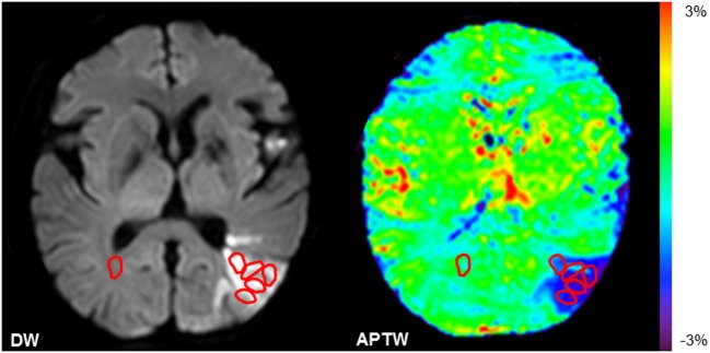
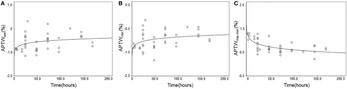
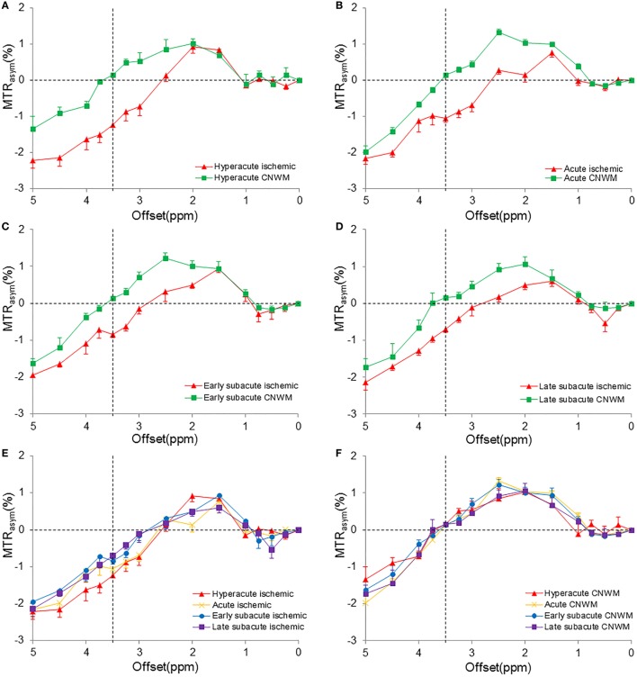
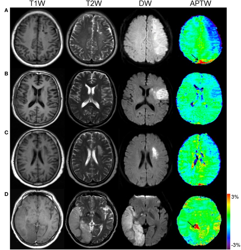
Amide proton transfer-weighted (APTW) magnetic resonance imaging (MRI) has recently become a potentially important tool for evaluating acidosis in ischemic stroke. The purpose of this study was to evaluate the dynamic pH-related changes in the lesions in patients with ischemia. Thirty-nine patients with ischemic stroke (symptom onset to imaging time ranging 2 h-7 days) were examined with a 3.0-T MRI system. Patients were divided into four groups: at the hyperacute stage (onset time ≤ 6 h), at the acute stage (6 h < onset time ≤ 48 h), at the early subacute stage (48 h < onset time ≤ 96 h), and at the late subacute stage (96 h < onset time ≤ 168 h). The APTW signal intensities were quantitatively measured in multiple ischemic regions for each patient. Compared with the contralateral normal white matter, APTW signals were significantly lower in ischemic tissue for all four stages ( < 0.05). The APTW signal intensities (APTW and APTW) increased consistently with onset time ( = 0.11, = 0.040; = 0.13, = 0.022, respectively). APTW showed a continued reduction with onset time ( = 0.44, < 0.001). Our results suggest that persistent tissue acidification could occur after ischemia, and as the time from stroke onset increases, the acidotic environment would alleviate. APTW signal intensities could reflect pH-weighted properties in ischemic tissue at different stages and time points.
酰胺质子转移加权(APTW)磁共振成像(MRI)最近已成为评估缺血性脑卒中酸中毒的一种潜在重要工具。本研究的目的是评估缺血患者病变中与pH值相关的动态变化。39例缺血性脑卒中患者(症状发作至成像时间为2小时至7天)接受了3.0-T MRI系统检查。患者分为四组:超急性期(发病时间≤6小时)、急性期(6小时<发病时间≤48小时)、早期亚急性期(48小时<发病时间≤96小时)和晚期亚急性期(96小时<发病时间≤168小时)。对每位患者的多个缺血区域进行APTW信号强度的定量测量。与对侧正常白质相比,所有四个阶段缺血组织中的APTW信号均显著降低(<0.05)。APTW信号强度(APTW和APTW)随发病时间持续增加(分别为=0.11,=0.040;=0.13,=0.022)。APTW随发病时间持续降低(=0.44,<0.001)。我们的结果表明,缺血后可能会发生持续性组织酸化,并且随着卒中发作时间的增加,酸中毒环境会得到缓解。APTW信号强度可以反映不同阶段和时间点缺血组织中的pH加权特性。